

Microscopes have opened up the small world to us. They have expanded every scientific field from biology to material science to ecology. The mechanism is pretty simple. We take something small and make it bigger! We expand it so that our vision can interpret the little details we normally couldn’t see. This is done by a light source and a series of lenses.
Light is sent to the object we wish to see – it bounces around and probes the object – it gets sent through some magnifying lenses – and then we see the enhanced image. Through the light and lenses, we create a magnified platform of which we can see (to a certain degree of magnification) the object and its environment much more clearly.
Field of View
Typically, microscopes are made up of several lenses and pathways for the light to travel to the object and eventually to our eyes.
To engineer and create these components, making them circular is the easiest and produces the highest quality images. This is why most microscopes have circular lenses and tubes and cylinders.
So when light travels through these, it makes sense that this micro “environment” we will see that is magnified will be a circle.
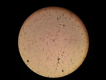
The magnified image is created! Scientists and engineers want to do more than just see stuff through the microscope. They want to probe the objects in it, measure certain dimensions. So they need a “ruler” to measure objects. But you can’t really… stick a ruler in this environment… you’ll mess up your sample!
However, because we know what kind of lens we are using (and thus the magnification they are providing), we can define microscope field of view. A microscope’s field of view is basically the diameter of that circular area that appears when you look into a microscope. Simple enough, right? We’ll look at some example and see how scientists and engineers calculate this and use this.
Examples of how to use Field of View
Any time you use a microscope, there should be one value that is of upmost important: the magnification of the lens you are using. Below we’ll see three examples of three different magnification lenses.

Every microscope’s eyepiece has its own magnification and also field number. There is a handy formula to relate all the numbers we have now: mainly the field number, field of view, and objective magnification:
Field of View (FoV) = (Field Number (FN))/(Objective Magnification)
Let’s say our Field Number is 50 millimeters. For the 5x magnification, we get a Field of View of 10 millimeters. For 10x we get 5 millimeters. For 50x we get 1 millimeter. We know have a length scale to apply to our microscope images!
Length scales with different magnifications:
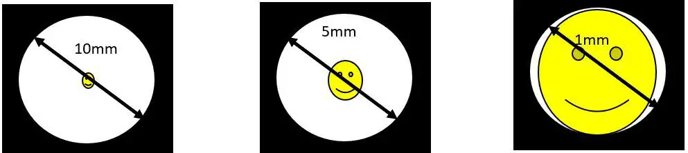
We can use this to measure objects in that field now! Let’s say we have multiple smiley faces in the first two magnifications:
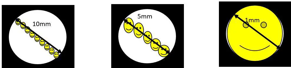
So, we can see from the first one that at a magnification of 5x, our field of view is 10mm, which is roughly 9 smiley faces. We can guess that one smiley is 0.9mm in diameter.
At a magnification of 10x, we have a field of view of 5mm, which is roughly 3 smiley faces. So, we can guess that each smiley is about 1mm in diameter. And finally, for the 50x, our smiley is slightly larger than the field of view, so we can guess that it is around 1.1 mm in diameter.
Different Fields of View for Different Applications
But isn’t smiley supposed to be the same diameter? Well – that is where some ambiguity in how microscopes function. The higher magnification, the more detail you get because you focus your field of view onto a smaller area. So, if you wish to study one and only one object (say, only one smiley face), then higher magnification is better. But maybe you want to study how one object interacts with another. You cannot use the highest magnification! You have to use a lens with a smaller field of vision a.k.a. smaller magnification.
That is where there are a ton of trade offs on which lenses to use and for when. If you want a very detailed measurement, higher magnification is needed. But if you wish to study two objects interacting with one another, then a lesser magnification is needed to capture both objects.
You must give up some information (i.e. the accurate measurements you can take at higher magnification) to earn some other information (i.e. the interactions between objects). A Scientist or engineer needs to pick and choose their field of view very carefully to fit their specific application.
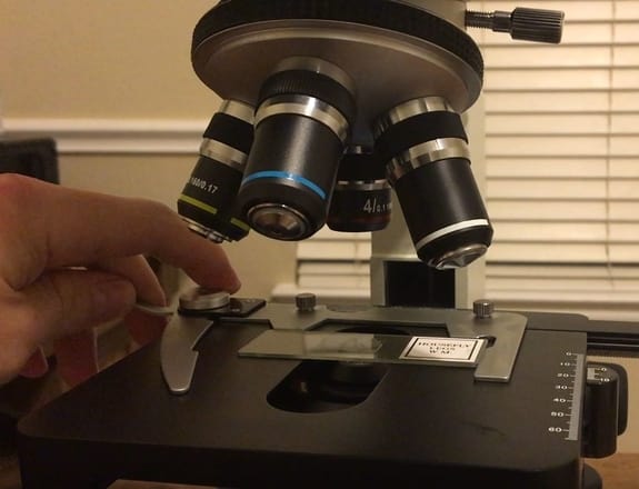
Changing Field of View: Different Lenses
Generally, every microscope is designed with two magnifications. It has one built into the top eyepiece. For more on the eyepiece see Parts of a Compound Microscope: Diagrams and Video.
This piece is built into the microscope. It has usually very small magnification (if any at all!) and no field number. The more important and higher magnification lenses are above the sample stage called objective lenses. For much more on objective lenses see Microscope Objective: The Eyes of the Microscope.
It is these lenses that provide most of the magnifying power to the light and to the image. To switch between different magnifications, they lens are usually mounted on a swivel. The user can rotate this swivel to easily switch from 5x to 10x or to whatever-x magnification they need.
By easily switching which lens is being used, the user and change the field of view very quickly – and thus see more or less detail and more or less of the magnified environment rapidly.
Takeaways
The key thing to remember here is that a microscope’s field of view is really the area that you can actually magnify and look at after the light is resolved in your eye. It depends on the magnification of the lens you are using, and another factor called the field number – which is related to the lens you are using.
At higher magnifications, you decrease your field of view at the expense of seeing things at higher details. At lower magnifications, you increase your field of view but lose some of the details you’d see at higher magnification. It all really depends on the application you are going for. But field of view is a driving parameter in how scientists and engineers use microscopes – and interpret what they are seeing.