
Rotifers are microorganisms that inhabit mainly freshwater aquatic environments and can range in size from 200 to 500 micrometers long. Rotifers are animals of the phylum Rotifera. They can be found mainly in freshwater within moist soils, still waters, and free-flowing waters. Rotifers have a unique crown of cilia around their mouth which allows them to create a vortex current which helps them pull in their food. Their characteristic foot and toe allow the Rotifer to stick to surfaces as well as move in a controlled manner.
Peering into a microscope to observe the Rotifers is comparable to peering into the past. Understanding the microscopic universe helps us better understand the roots of all life forms. Let’s look at what makes the Rotifer such a unique and special organism.
Common Types of Rotifers
Rotifers are abundant in species and are easily observed with a microscope. Unfortunately, the soft-bodied Rotifers do not fossilize well so there is not a robust fossil record of different species within Rotifera. Much of what we know about Rotifers are what we can observe today and infer into the past with our current knowledge.
Some of the most commonly known species of Rotifera are described below:
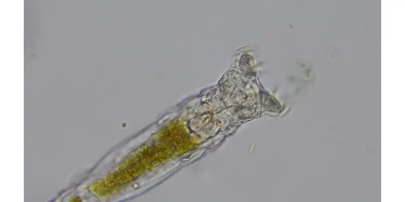
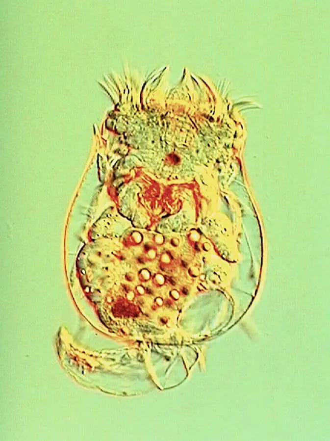
Brachionus calyciflorus: The Brachionus genus of rotifers occur mainly in freshwater and slightly brackish water. They can also be found in harsher aquatic environments such as alkaline. They are planktonic by nature, which means that they are unable to swim against the current and are mostly free-floating organisms. The calyciflorus are widely used as test animals in toxicology as they are particularly sensitive to pollutants. Their sensitivity also makes them great model organisms for other biological fields. The calyciflorus are found in large numbers and are often used as a zooplankton replacement at fish hatcheries, although don’t contain nearly enough nutrients to nourish fish larvae. There has been extensive research in genetically modifying this species to improve their lipid numbers, vitamin value, and mineral composition.
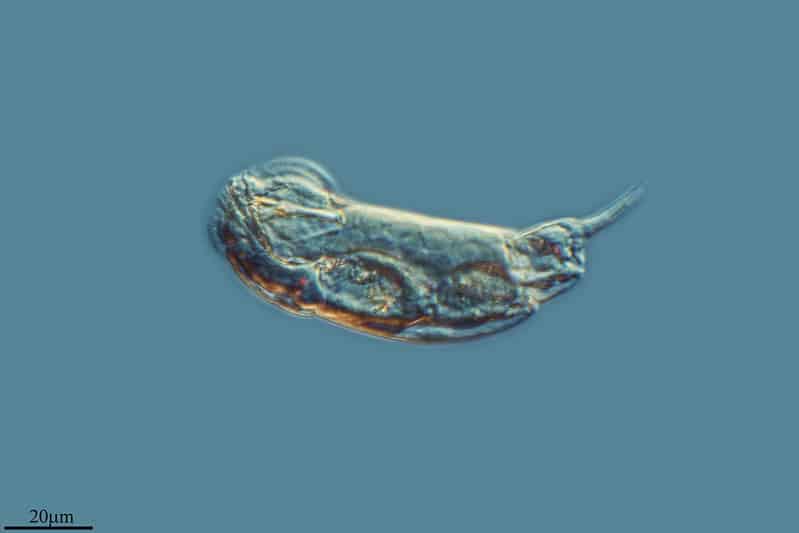
Colurella adriatica: Much like the calyciflorus, the adriatica are used as a zooplankton replacement at fish hatcheries. Unlike calyciflorus, the adriatica are much more tolerant to salinity and therefore used at marine fish hatcheries. They are not able to tolerate full-strength seawater for extended periods of time which is a good thing and a bad thing. Fish larvae typically consume all the adriatica before this becomes a problem. It’s a good thing as it helps preserve salt when storing and breeding the species. The species is flattened laterally and appears mussel-shaped when on its side.

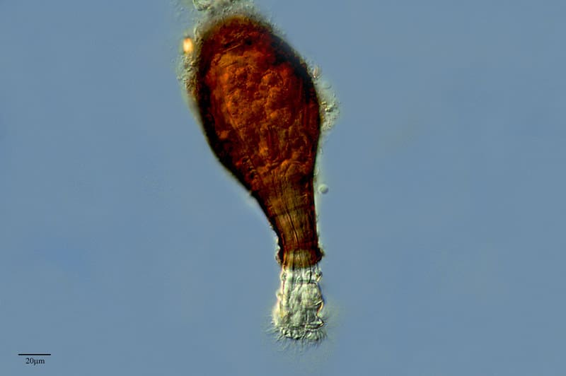
Habrotrocha angusticollis: The angusticollis species is one of the few species of Rotifers that has a hardened outside shell. Thanks to this unique feature, the angusticollis has an extensive fossil record. Fossils of the species are most commonly reported in peatlands and other habitats that contain moss. In particular, the species has been fossilized and recovered in northern Ontario. The angusticollis is easily recognized by its uninflated party balloon shape.
Cephalodella gibba: The gibba species are found in shallow and brackish waters. They are currently observed on the beaches of Australia, Central America, and Northern Europe. The gibba have the ability to move like a cephalopod and are within a genus of rotifers that are in the family Notommatidae.
What do Rotifers Eat?

Rotifers are detritivores in nature and eat decaying bacteria, algae, and protozoans. They can feed on organisms up to 10 micrometers large. Much like the crustaceans of the oceans, Rotifers are a benefit to the natural world by contributing to the recycling of nutrients. Because of this, cultures of Rotifers are oftentimes added to fish tanks or fish hatcheries to help improve the water quality. Rotifers may also be cannibalistic. As Rotifers contribute to nutrient recycling, they are often targeted by carnivorous secondary consumers such as shrimp and crabs.
Most species of Rotifer feed through their mouth. The corona, or the crown of the mouth, is lined with cilia. Cilia are short hairlike structures that vibrate and create a vortex of water that attracts food into its mouth. Rotifers then sift through the food particles and pass it through their digestive system. Because of its rather docile feeding mechanisms, Rotifers are unable to eat anything larger than what it can fit in their mouth.
What is the Structure of Rotifers?
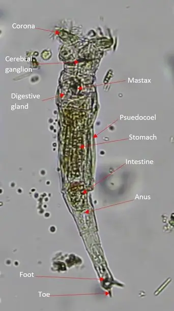
The structure and anatomy of Rotifers is not terribly complex. Let’s break it down below:
- Toe: The toe of the Rotifer has the ability to attach itself to objects. It uses a cement gland to excrete a sticky substance that allows the Rotifer to latch on. While attached, the Rotifer can relax and sift. Rotifer sometimes attaches themselves to crustaceans or other mobile organisms for a free ride to sources of food. John Harris compared the toe of the Rotifer to ‘forceps’ in his letter.
- Foot: The foot is present mainly in sessile Rotifers and is virtually nonexistent in free-swimming species of Rotifers. The foot consists of up to four toes. It acts as a support to the toes and is more comparable to an ankle, allowing the species to rotate but also giving it support and stability.
- Anus: After food is digested and passes through the intestines, the waste product is excreted through the anus. In some species, the waste product is excreted through the trophi instead.
- Intestine: The intestine is short and terminates in a posterior orifice called the cloaca that acts as a urinary tract. It has the ability to excrete both urine and feces.
- Stomach: The stomach of the Rotifer is directly after the chewing pharynx and is covered in ridges. This is where the digestion of food and absorption of nutrients occurs. A total of seven salivary glands exists which includes two gastric salivary glands that help produce digestive enzymes.
- Mastax: Also known as a pharynx, the mastax is a ciliated tube with muscles and hardened jaw-like structures called trophi that help chew and grind food as it passes through the esophagus. The mastax ensures that food is properly smashed and broken down before it can pass through to the stomach for digestion and absorption.
- Mouth: The mouth does not have the ability to unhinge or open any wider. It is also not capable of closing. It’s simply a cylindrical opening that allows for food to pass through.
- Corona: The corona is a defining characteristic amongst the Rotifers and its shape and size vary greatly from species to species. Different shapes correlate with different genus, although the shape should not be the only characteristic used to identify a Rotifer genus. For a majority of Rotifers, the corona has two different ciliated rings called the trochus and cingulum. Each structure helps create an inward water current. These structures can also create enough current to swim freely (in some species). The inward current is used to bring food into the Rotifers mouth to feed on. In some rare cases, the ciliated corona is completely absent. Depending on the species, there may also be sensory antennae, pals, cirri, or vestibulum present as well.
- Pseudocoel: This body cavity is analogous to a coelom but is not lined with mesodermal cells. It’s a fluid-filled cavity that helps suspend the digestive system within the body. It plays no role in gastrulation.
- Vitellarium: The vitellarium is a type of gland that secretes yolk that turns into eggshells. It secretes a protein that hardens called sclerotin.
The trophi and the gut are defining characteristics of the Rotifer’s digestive system. The trophi are made up of chitinous material that appears much like a jaw. Food sources are either pierced or smashed up before they can pass through to the stomach. Some species of Rotifers use this structure as a food-storage organ, although most species choose to digest the food in their stomach.
Each Rotifer has a complex muscular system that comprises longitudinal muscles as well as circular muscles that each have their own unique function. Muscle contraction as well as the fluid-filled pseudocoel act as the Rotifers skeleton and give it its rigidity. Muscles of the Rotifer are also responsible for retracting its food and controlling the movement of its appendages. Some arm-like structures of the Rotifer are capable of deterring predators by quickly swinging them outwards. The neural system of the Rotifers is controlled at the cerebral ganglion.
It connects to sensory organs that can identify variations in pressure, light, or toxins. Some organs consist of receptors of mechanics, chemicals, and more. The mechanoreceptors and chemoreceptors are found within the corona whereas the photoreceptors are located at the eyespot of the organism near its head. The receptors work in tangent to notify the Rotifer of any danger or to help move them to a more favorable environment.
How are Rotifers Classified?
All living organisms are classified based on a rated system of related species based on structure and genetics. The Domain is the uppermost tier of the ranking system, followed by Kingdom, Phylum, Class, Order, Family, Genus, and Species. Rotifers belong to the Phylum Rotifera and can further be broken down from there.
Let’s examine how Rotifers are classified:
- Domain: Eukaryote – In a three-domain system, Rotifers fall within the Eukaryotes. Each eukaryote has a nucleus that contains important genetic material that contains hereditary information that is encased by a nuclear membrane. Eukaryotes may be multicellular or unicellular. Animals, plants, protists, and fungi are grouped within this domain. Organelles like the mitochondria and Golgi apparatus are all membrane-bound.
- Kingdom: Animalia – A group of multicellular organisms that consume organic materials, breathe oxygen, are mobile, can produce sexually, and grow through a process of embryonic development are known as the Animalia. 1.5 million species and counting are within this Kingdom, including about a million species of insects. It’s estimated that there are around seven million species within the Kingdom in total as many have yet to be discovered and characterized.
- Subkingdom: Eumetazoa – Otherwise known as diploblasts, this proposed clade is a sister group to Porifera. Identifying characteristics of eumetazoans include true tissues arranged into germ layers that take shape during embryonic development, the existence of neurons, and an embryo the undergoes gastrulation.
- Phylum: Rotifera – Many of the rotifers are freshwater organisms that are free-swimming and planktonic, meaning they are unable to propel themselves against the forces of current. With that known, Rotifera are most commonly found in still water or within moisture drops of moss and soils. Rotifera have a cosmopolitan distribution, meaning that their characteristics are extremely similar from species to species across the globe.
- Class: Seisonidea – A small family of rotifers that live on the gills of small crustaceans within the genus Nebalia. This class is unique because the males and females are the same sizes, which is uncommon amongst Rotifers. They are thought to be close ancestors to early species of Rotifers and have a long body with a reduced corona. Seisonidea have a muscular complex that is identical to other Rotifers, which includes longitudinal muscles and striated rectus muscles.
- Class: Bdelloidea – With over 450 documented species, the bdelloidea are distinguished entirely on their morphological differences. Parthogenic reproduction and their unique ability to survive through unfavorable conditions such as dry weather are unique to the bdelloidea. They have the ability to enter a form of dormancy to protect themselves at any point of their development. This unique asexual history dates back over some 25 million years ago through fossil evidence, so they are often referred to as the ‘ancient asexuals’ of the Rotifers.
- Class:Monogononta – The largest group of Rotifera with over 1500 species known to mankind. This class includes both free-swimming and sessile (the lack of locomotion) forms of Rotifer. This class has a reduced corona and typically only one gonad, which is where the name monogononta comes from. Males are typically smaller than females and the two produce seasonally. Some females have the ability to reproduce through parthenogenesis, although uncommon.
Where do Rotifers Live?

The great majority of Rotifer species live within freshwater environments. Since they are planktonic, meaning that many don’t have the ability to swim against the current, Rotifers prefer still waters like lakes and ponds. They can also be found within water films of mosses, leaves, soils, and lichens. Most species are freshwater although some can tolerate brackish to salty environments.
The most common marine Rotifers choose to hitch a ride and live on other marine crustaceans. This way, they don’t have to fight against the current of the ocean themselves and are transported to other food sources without having to use up any energy of their own. In rare cases, Rotifers have been found as deep as 400 meters in the ocean. Around 25 species of Rotifers are free-swimming, and many are colony members. On rare occasions, rotifers can be parasitic in nature.
Since Rotifers are considered cosmopolitan species, meaning their characteristics don’t greatly vary from creature to creature across many different environments across the globe, they are an intriguing study as they flourish despite ecological barriers. Environments that Rotifers have been found in include arctic waters, hot springs, acidic waters, saltwater, temporary water bodies like puddles, terrestrial environments, and more. Others can be found in open water in planktonic forms, sediments, and even within other organisms.
How do Rotifers Reproduce?
Rotifers can reproduce in a number of different ways depending on their species, environment, and even diet. Common forms of reproduction are cyclical parthenogenesis and amphoteric reproduction. The short lifespan of 40 days means that the ability to reproduce under virtually any circumstance is vital to the species’ survival.
In fact, reproduction has become so mainlined and efficient in Rotifers that some species only have females and rely entirely upon parthenogenesis. This is uncommon as most species have both males and females within their population. It’s unique that some species of rotifers have opted throughout evolution to focus solely on asexual reproduction.
Cyclical parthenogenesis does not require a male to help fertilize an egg. Instead, the females can produce their own diploidal egg. Depending on the species, more than one egg can be produced throughout the year. The eggs then undergo mitotic division which is a form of division in eukaryotic cells when a parent cell divides into two identical daughter cells.
Some species of Rotifers have males and sexual reproduction is possible. This is contingent upon the female of the same species producing mictic offspring that are even capable of sexual reproduction. Amictic offspring, or offspring that are incapable of sexual reproduction, continue to be produced despite males being present. This helps keep the proportions of the group in balance and is entirely dependent upon water temperature, chemicals in the water, and more.
In instances where male Rotifers and capable female Rotifers are in the same colony, mating events occur in which the male fertilizes the female’s eggs to produce an embryo. If for whatever reason, the eggs are not fertilized, then the egg continues to produce haploid male offspring.
Fertilized eggs are diploidal and produce eggs with protective walls that encase them. The protective shells are capable of withstanding unfavorable environmental conditions. When conditions become favorable, the eggs hatch and give life to amictic females that reproduce asexually.
Amphoteric reproduction occurs where females can give life to both males and females. Males are born through haploid eggs whereas females are born through diploid eggs. As you can see, Rotifers are extremely unique in how they reproduce. Because of this, Rotifer are highly distinguishable by their reproductive tendencies and also widely studied thanks to their asexual preferences. Although not particularly resilient in unfavorable conditions, the eggs of Rotifers are.
Are Rotifers Harmful to Humans?
Rotifers are not harmful to humans. To date there are no known human diseases caused by ingesting or coming into contact with Rotifers. In fact, Rotifers may be the key to stopping a parasitic worm that is harmful to humans known as a schistosome or blood flukes. Schistosomiasis is a disease caused by the schistosome entering the body and the body’s reaction to its eggs.
Curiously schistosome have been known to be paralyzed by a substance apparently emitted by rotifers. When scientists observed the parasitic worms in the presence of Rotifers or even in water once occupied by Rotifers the worms were in a frozen immobile state. This observed phenomenon has prompted further study into the application of Rotifers in fighting Schistosomiasis.
How to Observe Rotifers Under a Microscope
Rotifers are one of my favorite microorganisms to observe under the microscope. They truly look like something out of a science fiction novel. It is speculated that Rotifers were at least in part the inspiration for the look of the “Demogorgon” creature in the popular Netflix series “Stranger Things”. Rotifers are very common and can be seen sticking its toe to just about any surface and slinking its way across the field of view. You can see most Rotifers easily under 100X magnification but if you can get one that has stopped in one place you can see some great detail of the toe and corona at 600X magnification.
Rotifers are extremely common but if you are gathering a sample, I would recommend making sure you get some algae or sediments which should contain quite a few. When Rotifers stop to open up the corona you can get a great look at the vortex that is created and see all the little cilia that make that possible. Rotifers have so many detailed amazing features that make them really fun to observe and are great for sparking interest in the microscopic world.
When Were Rotifers First Discovered?
There are two contenders who first discovered the Rotifers. The first contender is Antoine van Leeuwenhoek who is widely recognized as the father of microbiology. The second contender is John Harris, an English writer, scientist, and priest.
In October of 1687, Leeuwenhoek sent a letter to the Royal Society that contained what is believed to be the first description of the Rotifer. The letter included a crop of the root of duckweed which depicted the organisms attached to it. A rotifer is meticulously depicted in two separate positions.
Another letter of Leeuwenhoek’s was sent to Hendrik van Bleyswijk with even more clear illustrations of the Rotifer. The letter that was sent in 1702 showed a rotifer when fully extended and even labeled different body parts. The species was assigned Philodina roseola at the time.
Others believe that it was John Harris who first wrote of the Rotifer in 1694. Harris was best known for his work as the editor of Lexicon Technicum: A Universal English Dictionary of Arts and Sciences. Harris later served as the vice-president of the Royal Society and worked as a mathematics professor.
John Harris’ 1694 letter recounted an observation of standing rainwater in a pot on his windowsill. He explained the organism that he witnessed as being similar to a maggot that has the ability to contract and stretch. He also described a tail that looks like a pair of forceps. Although vague, many scientists believe that the described creature very well may be a Rotifer.
Historians have trouble agreeing on who first identified the Rotifer due to language barriers and translations. Much of Leeuwenhoek’s work was lost to translation or not interpreted until long after it was written. Additionally, the sheer volume of letters that Leeuwenhoek sent out describing a multitude of microorganisms makes the history of it difficult to follow.
References
- https://www.arcella.nl/habrotrocha-angusticollis/
- https://onlinelibrary.wiley.com/doi/abs/10.1111/j.1365-2095.2007.00504.x
- http://www.microscopy-uk.org.uk/mag/indexmag.html?http://www.microscopy-uk.org.uk/mag/artoct15/dw-snippet-2.html
- https://link.springer.com/chapter/10.1007%2F978-94-009-1583-1_35
- https://www.risingtideconservation.org/colurella-adriatica-update/
- https://ucmp.berkeley.edu/phyla/rotifera/rotifera.html
- https://animaldiversity.org/accounts/Rotifera/