
Bodies of animals, plants, bacteria, and fungi are all made of cells. The cells of various groups of organisms may vary significantly from each other. However, the various cells of the same kind of animal or plant, and even inside the same organism can belong to different types of tissues which are made up of different kinds of cells. There are epithelial, connective, muscular, neuronal tissues etc. Organs in the body are made of the cells of different tissues which work together to allow organs and, ultimately, the entire body to be alive and active.
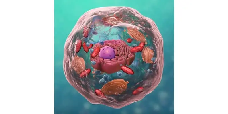
Organelles are just organs within the cell. The same way the human body has organs, where each organ performs a very specific task and all organs work together to keep the body functioning and thriving, so too do cell organelles within a cell.
The cell is a smallest structural unit of living organisms (1-4 micrometers). How small are they? The biggest human cell is an ovum. An ovum is a female reproductive cell and measures 100 micrometers in diameter (one tenth of a millimeter).
Most human and animal cells are usually smaller, around 10-20 micrometers or less. For example, the diameter of white blood cells is between 7-10 micrometers. So, when we are talking about cell organelles are talking about extremely small structures. In this post we will explore some common cell organelles and go through their function and role in making the cell an efficient system.
Cell Organelles
The word “organelle” means “little organ”. Like organs inside the animal or human body, organelles are located inside the cell. It is important to distinguish between parts of the cell that are classified as organelles and those that are not. The animal cell organelles include:
- Endoplasmic reticulum.
- Ribosomes.
- Golgi apparatus.
- Mitochondria.
- Endosomes.
- Lysosomes.
- Peroxisomes.
- Nucleus.
There are many other parts of the cell that are very important to the overall structure and functioning of a cell but are not considered technically organelles. Some examples are the plasma membrane and components of the cytoskeleton like tubulin microtubules, actin filaments (microfilaments), and intermediate filaments.
Endoplasmic Reticulum
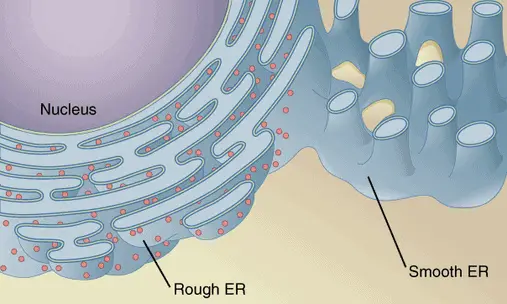
Approximately one half of all membranes in the cell belongs to the organelle called the endoplasmic reticulum (ER). There are other organelles called ribosomes that are located on the surface of the endoplasmic reticulum. The rough or granular endoplasmic reticulum is responsible for the synthesis of proteins. There is also smooth or agranular endoplasmic reticulum which has no or almost no ribosomes on its surface. The endoplasmic reticulum is also where lipids are synthesized for all the cell.
The endoplasmic reticulum is a central organelle which plays an important role in the transport of biomolecules like proteins. In this process the endoplasmic reticulum collaborates with Golgi apparatus. For example, digestive enzymes produced in the secretory cells of pancreatic gland are transported by the endoplasmic reticulum and then by Golgi apparatus from the site of protein formation (from ribosomes located on the rough endoplasmic reticulum or in cytosol) to the site of Golgi apparatus where secretory vesicles form.
The following transportation steps are done by secretory vesicles that move the enzymes and other proteins through cytoplasm to various intracellular locations or to the plasma membrane where biomolecules are excreted outside the cell by merging of vesicles with the membrane.
The rough and smooth endoplasmic reticulum are also non-technically referred to as “factories” for enzyme-powered synthesis of proteins and lipids. As you know, proteins and lipids are building materials for the body. Of course, all organelles and other structures in the cell are made of proteins and lipids, too. Both endoplasmic reticula have a large surface area of its membrane where plenty of enzymes localize and participate in the production of proteins and lipids.
Proteins synthesized by ribosomes in cytosol (not in the granular endoplasmic reticulum), came into the endoplasmic reticulum due to an activity of the special signal peptide which mediates their transfer from the cytosol into the endoplasmic reticulum.
Ribosomes
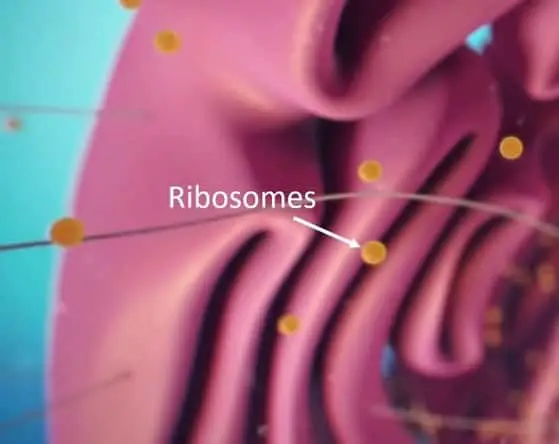
Ribosomes are organelles which are responsible for the synthesis of complex membrane proteins and soluble proteins which are needed for the building of or the use by other organelles or for secretion outside the cell. They provide their service locating in the cytosol or being attached on the membrane of rough or granular endoplasmic reticulum.
Ribosomes are made of proteins and ribosomal ribonucleic acid (rRNA). Ribosomes link amino acids together. Each ribosome comprises two subunits. Messenger RNA (mRNA) comes from the nucleus, bringing genetic information from the DNA, and binds the smaller subunit of the organelle.
The second bigger subunit of the ribosome is responsible for attaching the amino acids to the growing chain of the building protein. This is performed by the rRNA. Amino acids are transported from the cytosol to the ribosome by the transport RNA (tRNA). As you can see, three types of RNA are used during protein synthesis by ribosomes.
Golgi Apparatus
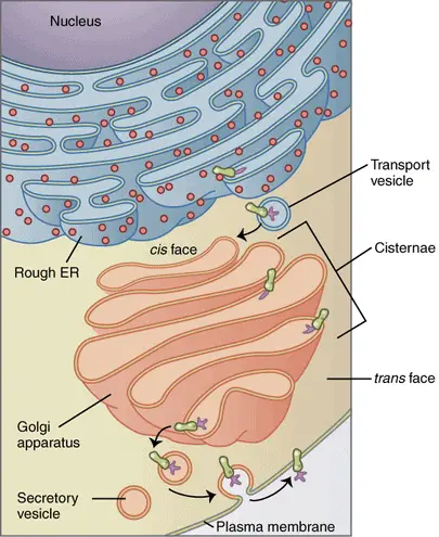
Golgi apparatus (also called Golgi complex or Golgi body) is an organelle network of membranes, fibrils and granules. It is usually located near the cell nucleus and often near the centrosome.
The Golgi apparatus has two surfaces: trans-surface mature and cis-surface developing membranes. The Cis-surface of the Golgi complex is tightly linked with endoplasmic reticulum. Trans-surface of the Golgi complex produces a network (reticulum) of tubes called Golgi network. Golgi apparatus is built of flattened membrane tanks (called Golgi cisternae) resembling a stack of plates. Each such stack is made of 4-6 cisternae with a diameter around 1 micrometer.
The cell may have one large or multiple small stacks of Golgi cisternae. Other structures belonging to the Golgi complex are tiny membrane bubbles (called Golgi bubbles) located near Golgi cisternae. Their role is the transportation of proteins and lipids between Golgi cisternae and from Golgi apparatus to various intracellular locations. The Golgi apparatus receives a variety of proteins and lipids from the endoplasmic reticulum, modifies, and transports these biomolecules to various sites inside the cell.
The Golgi apparatus is responsible for assembly and modification of the complex proteins called proteoglycans by attaching sugars and other molecular components, in the presence of special enzymes, to the side chains of proteins. Some proteoglycans are secreted by the cell as a part of a protective mucus, others are incorporated into the structure of the plasma membrane.
Mitochondria
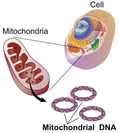
Mitochondrion (plural of mitochondria) is an organelle with a bean-like body composed of a double membrane. Mitochondria measures 0.5-1 micrometers in diameter and up to 7 micrometers in length. The cell may contain 2000 mitochondria that occupy up to 25% of the cell’s volume! There is also a correlation between a lower number of mitochondria in the cell and a larger size of cell organelles.
Mitochondria have their own genome (own DNA) and even their own ribosomes! Interestingly, mitochondrial proteins are encoded by both genomes. Meaning the proteins are encoded by the genome of the mitochondrion and the genome of the whole cell.
The mitochondrion contains four compartments: matrix, inner rough membrane, intermembrane space, and outer smooth membrane. The membranes are separated by intermembrane space. The inner membrane of a mitochondrion has folds called cristae. Presence of cristae in mitochondrion is a structural advantage which aids aerobic cellular respiration.
The function of mitochondria is a production of universal energy for the cell. The mitochondrion is, in non-technical language, a “power plant” of the cell.
The organelle employs two complex biochemical processes (electron transport and oxidative phosphorylation) to produce the special biomolecule called АТР (adenosine triphosphate). The adenosine triphosphate molecule is used for generating and storing of the biological energy which is consumed by other cellular organelles for all cellular needs and activities. To produce adenosine triphosphate, the enzyme adenosine triphosphate synthase and a variety of cytochromes localize and work in the cristae of mitochondria.
Transport proteins and enzymes of the respiration chain are found in the mitochondrion’s matrix. Inner and outer mitochondrial membranes are permeable to oxygen, carbon dioxide, and water. While the outer membrane is more permeable to ions and small molecules, the inner membrane creates compartments by separating the matrix of mitochondrion from the cytosol.
Mitochondria alongside with endoplasmic reticulum is also a storage organelle (a depot) for calcium ions. Mitochondria and the endoplasmic reticulum maintain a constant low level of these ions in cytosol and release them during cellular activation. Such regulation of calcium metabolism is extremely important in muscle contraction and expansion.
Endosomes
Endosomes, non-technically referred to as recycling compartments, are organelles of animal cells which represent membrane-bound vesicles responsible for endocytosis. Endocytosis is the process of ingestion of molecules and particles by the cell. Endocytosis allows surface molecular receptors of the cells to be recycled and expressed again in a new place on the cell surface.
The endocytosis process is a remarkably important process in the activities of immune cells, participating in both the innate and natural immunity (phagocytic activity of pathogens by macrophages) and acquired and active immunity (interaction of T cells with antigen-presenting cells like macrophages, B-cells, dendritic cells and the presentation of antigens to T cells). Endocytosis is the reverse process of exocytosis (cellular secretion) conducted by the transport vesicles which we discussed above. Prokaryotic cells have no endosomes (as well other organelles) and they do not undergo endocytosis and exocytosis.
Lysosomes
Lysosomes are organelles that contain digestive enzymes which degrade dysfunctional and/or old organelles. Lysosomes also utilize various particles and molecules ingested by the cell in the process of endocytosis conducted by organelle endosome (as mentioned above). The lysosomes are small (0.2-2 micrometers) but the cell may have up to several hundreds of those organelles.
There are about 40 types of digestive enzymes (called hydrolases) in the lysosomes. While cytosol has a neutral pH (~7-7.3), the inner media of the lysosome has the acidic pH of 4.5-5, which is found to be an optimal condition for many lysosomal enzymes. Hydrolases of lysosomes are inactive at the cytosol where pH is different. Lysosomes and their role in the breakdown of particles, pathogens, toxins are also important in immune processes of phagocytosis, non-specific and specific immune cytotoxicity, interaction of T cells with antigen-presenting cells, and antigen presentation.
Lysosomes are also important in apoptosis or “the cell suicide” when the cell destroys itself to prevent or minimize a bigger harm to the whole body.
Peroxisomes
Peroxisomes, also called microbodies, are small spherical organelles enclosed by a single membrane. Their diameter in most cells usually ranges from 0.15 to 0.25 micrometers, but in the liver cells, peroxisomes are much larger (from 0.5 to 1.5 micrometers). While the peroxisomes do not have their own genome (unlike mitochondria which do), they are still capable to replicate in the cell by using genetic information stored in the cell’s genome. Peroxisomes are present in all eukaryotic cells.
However, the functions of these organelles can be different in the cells of various types of tissues. It is believed that peroxisomes and mitochondria have a unique membrane receptor which mediates the selective transport of proteins into these organelles from cytosol. Peroxisomes, like mitochondria, are responsible for utilization of oxygen in the cell. Peroxisomes also produce toxic intermediates from oxygen, for example, oxygen peroxide (Н2О2), which they employ for oxidation. By the way, the name of peroxisome is given due to enzymatic ability of the organelle to produce hydrogen peroxide.
Peroxisomes also generate catalase, an enzyme which they use to eliminate an excess of oxygen peroxide in the cell. In the liver cells, peroxisomes produce three enzymes: 2 oxidases and a catalase. Peroxisomes, particularly in the cells of liver and kidneys, use hydrogen peroxide to oxidize many toxic substrates like phenol, formaldehyde, alcohol etc. to neutralize the toxic activity of these substances for the cells and protect the whole body.
Nucleus
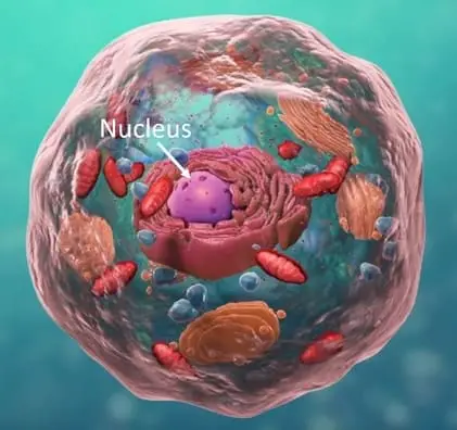
The nucleus can be thought of as the control center of the cell primarily because it controls and regulates cell activities like growth, metabolism, protein synthesis, and reproduction. The nucleus also contains genetic material called DNA.
DNA is made up of molecules called nucleotides. The nucleotide itself is made of the following material: a phosphate group, a sugar group, and a nitrogen base. Within the nitrogen base are the familiar nucleobases: adenine (A), thymine (T), guanine (G) and cytosine (C). If you’ve seen the part in Jurassic Park where they are watching the DNA presentation and the letters are flying by this is what they are talking about. DNA can be though of as a kind of biological source code that tells the cell what to do.
Nucleolus is a structure in the nucleus where ribosomes are made. The number of nucleoli within a nucleus will depend on the species but the number within a species is constant. The nucleolus is formed after cell division is complete and chromosomes have gathered and disappears again during cell division.
The nuclear envelope is analogous to an actual envelope in that is encloses the nucleus and its contents. Although the envelope contains the nucleus it does have tiny holes called nuclear pores which allow different molecules to pass between the nucleus and cytoplasm.
Evolutionary Considerations Related to Organelles
Mitochondrial ribosomes in eukaryotic cells are produced with the use of genetic information delivered by mRNA from the mitochondrial genome. These eukaryotic ribosomes functionally and morphologically resemble prokaryotic ribosomes of bacterial cells. This observation provided researchers with a suggestion about the evolutionary origin of mitochondria from bacteria.
In various experiments results showed that this evolutionary scenario could have taken place. For example, in an experimental study where there was an injection of nitrogen-fixing bacteria into host plant cells, the inserted bacteria formed new nitrogen-fixing organelles inside the plant cells. The bacterial cells were able to develop a symbiotic (partnership-like) relationship with the organelles and other structures of the eukaryotic cells. It is very likely, that similar processes were possible in the course of evolution of eukaryotic cells throughout hundreds of thousands of years.
During evolution of eukaryotic cells, some organelles could lose their functions or lose their importance in the cellular life and activities. For example, it has been suggested that peroxisomes of modern organisms originated from ancient organelles which were necessary to protect the cells from oxygen’s toxicity. Ancient peroxisomes decreased oxygen concentration in the cells and used oxygen’s activity in oxidative reactions. Appearance of mitochondria and the reaction of oxidation phosphorylation made the role of peroxisomes, which utilize oxygen without production of energy in the form of ATP molecules, less significant in modern organisms.
Can Organelles be Seen Under the Microscope?
The short answer is it depends on the microscope. The majority of organelles with the exception of the nucleus are actually smaller than the wavelength of the visible light spectrum making them impossible to see under a light microscope.
They are, however, visible under an electron microscope. For more on electron microscopes and how they work see Electron Microscopes Explained: From Physics to Images. That said there are some cases where the nucleus can actually be visible under a light microscope.
There are also other structural and non-organelle components like cytoplasm and the cell membrane that can be visible under a light microscope.
Relevant History
The first “real” compound microscope, which combined an objective lens with an eyepiece to view a magnified image, was invented, built and employed in the Netherlands by Antonie van Leeuwenhoek, the businessman and scientist, in the late 17th century. He made optical microscopes with a single lens and used them to conduct the first observations of bacterial cells of and protozoan cells. For a complete history or microscopes check out The First Microscope to Modern Microscopes: Evolution and History of Microscopes.
The wide availability of microscope in the beginning of the 19th century resulted in the high progress in the biological studies, particularly in the research focused on plant and animal cells and tissues.
In 1665, in England, a polymath, naturalist and architect Robert Hooke discovered and described the cell in a microscopy study conducted on a fruit fly’s eye and a plant cell.
In 1838, the German physician and physiologist Theodor Schwann and the German botanist Matthias Jakob Schleiden proposed the cell theory for animal and plant organisms, respectively. However, optical microscopes have limitations. The light microscopes cannot focus on objects with the size smaller than the wavelength of visible light.
In the beginning of the 20th century, scientists needed more powerful devices to look at organelles and study their morphology and physiology in detail. The first electron microscope was invented in the 1920s, but a more advanced model was built in Berlin, Germany in 1931 by the physicist Ernst Ruska and the engineer Max Knoll.
While many organelles of the cell have been discovered with the use of optical microscopes 19th century and the early 20th century, much smaller organelles ribosomes were first observed by the cell biologist George Emil Palade under the electron microscope in the mid-1950s in USA.
More Facts about Cells and Cell Organelles
The cell is a hallmark of life. For example, viruses are not made of cells, and cannot be considered as a regular form of life. However, even viruses could not exist without living cells of organisms! Viruses are specialized parasites and they cannot complete their life-cycle without infecting a suitable living host. Each group of viruses target the cells of a particular type where they can reproduce their genome and protein envelope by using the DNA, RNA, and protein-synthesizing machinery of infected cells.
Both animal and plant cells have mitochondria. However, plant cells have also other organelles (various colorful plastids including chloroplasts, chromoplasts, leukoplasts, geroplasts, vacuoles and structures like the cell wall.
Chloroplasts (green plastids) are amazing organelles found in the cells of green plants and algae. They have green pigment called chlorophyll which allow them to use the energy of sunlight to produce sugar and oxygen from carbon dioxide and water!
This process is called photosynthesis, a process which feeds all animals and humans! Animal cells are not capable of photosynthesis! Chloroplasts produce a major amount of ATP (adenosine triphosphate) like mitochondria in animal cells. This energy is consumed by chloroplasts in their biosynthetic reactions and other organelles of the plant cells.
Plant cells also have vacuoles, the organelles which look like large intracellular bubbles filled up with water, sugar and other nutrients. These storage facilities allow the plant cells to accumulate and save nutrients like sugar (obtained from photosynthesis) and water.
Vacuoles are also found in animal cells; however, they are much larger in plant cells. Vacuoles allow the plan cells to keep its structure by maintaining the osmotic (turgor) pressure in the cell against the cell wall. The cell wall is a structure made mostly of cellulose and also found only in the plant cells.
Takeaways
Just like the human body could not live without our organs, the same is true for these little organs we call organelles. These highly complex and very small organic machines perform complex processes at the subcellular level that makes life possible for the majority of visible biomass we see on the planet.
As amazing as the organs in our body are, you have to marvel at the sheer complexity and precision at which these organelles operate. I hope this has given you a good primer on cell organelles and will further your microbiological knowledge.
References
- The American Heritage Dictionary of the English Language. Second Edition. Boston: Houghton Mifflin, 1982.
- Bruce Alberts, Alexander Johnson, Julian Lewis, Martin Raff, Keith Roberts, and Peter Walter. Molecular Biology of the Cell. New York :Garland Pub., 2002.
- Lodish H., Berk A., Matsudaira P., Kaiser C.A., Krieger M., Scott M. P., Zipursky L., Darnell J. Molecular Cell Biology 5th Edition. New York: W. H. Freeman, 2003.
- Koolman, J., Röhm, K.H Color Atlas of Biochemistry. Thieme, 2011.
- Hassel J. R.. Kimura J. H., Hascal V. C. Proteoglycan core protein families Annu Rev. Biochem., 55, 539-567, 1986. https://www.annualreviews.org/doi/10.1146/annurev.bi.55.070186.002543
- Bailey, F. R., Copenhaver, W. M., Kelly, D. E., & Wood, R. L. Bailey’s textbook of histology. Baltimore: Williams & Wilkins, 1978
- Kaur G, Lakkaraju A. Early Endosome Morphology in Health and Disease. Adv Exp Med Biol. 2018;1074:335–343. https://www.ncbi.nlm.nih.gov/pubmed/29721961
- Coba de la Peña, Teodoro et al. The Symbiosome: Legume and Rhizobia Co-evolution toward a Nitrogen-Fixing Organelle? Frontiers in plant science vol. 8 2229. 22 Jan. 2018 https://www.ncbi.nlm.nih.gov/pmc/articles/PMC5786577/
- Atti Della Fondazione Giorgio Ronchi E Contributi Dell’Istituto Nazionale Di Ottica, Volume 30, La Fondazione-1975, p. 554
- Observationes microscopicae Antonii Lewenhoeck, circa particulas liquorum globosa et animalia. Acta Eruditorum. Leipzig. 1682. p. 321.
- Mazzarello, P. A unifying concept: the history of cell theory. Nature Cell Biology. 1999 1(1): E13–5,. https://www.nature.com/articles/ncb0599_E13
- Mathys, Daniel, Zentrum für Mikroskopie, University of Basel: Die Entwicklung der Elektronenmikroskopie vom Bild über die Analyse zum Nanolabor.
- Palade G. E. A small particulate component of the cytoplasm. The Journal of biophysical and biochemical cytology. 1955 1(1), 59–68. https://www.ncbi.nlm.nih.gov/pmc/articles/PMC2223592/
- https://www.ncbi.nlm.nih.gov/pmc/articles/PMC4130368/