
The Golgi apparatus is a cell organelle that receives newly synthesize proteins and lipids and is in charge of transporting them to different parts of the cell. If the nucleus can be thought of as the CEO or boss of the cell, the Golgi apparatus can be thought of the shipping department sending packages of protein to their destination.
If you’re not sure exactly what that means don’t worry. In this post we will go through, in detail, the primary functions, structure and the history of the Golgi apparatus and distill only the what you need to know to fully understand this very important organelle.
Golgi Apparatus Structure
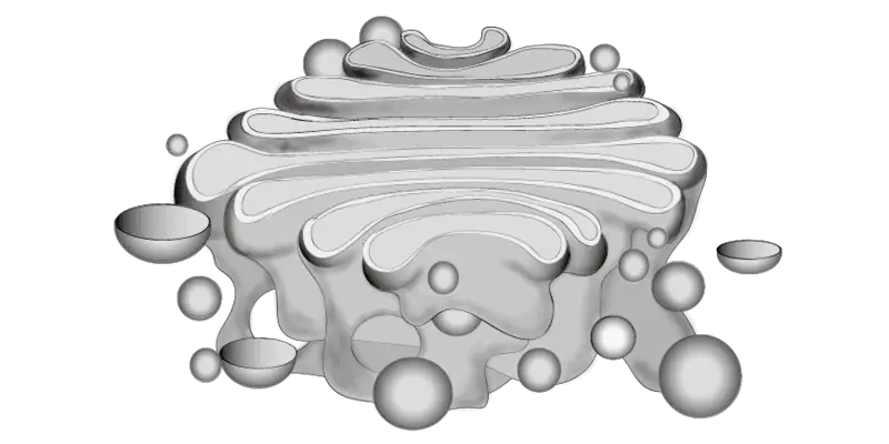
The Golgi apparatus is an organelle found in most eukaryotic cells that constitutes a complex system of stacks of cisternae arranged in a side by side fashion, tubules and vesicles, which are generally located in a special region of the cytoplasm known as the Golgi apparatus zone.
The distribution of the cisternae stacks may be spread more or less evenly in the cytoplasm in the case of plants and some animal cells. The Golgi apparatus is also made up of various vesicles and tubules. Although you may see standard textbook renderings of the Golgi apparatus, the morphological features of this organelle vary from one cell type to another, and even among the same types of cells (Morré & Mollenhauer 2008).
Cisternae by definition is a sac or a cavity usually filled with fluid. This term was initially used to describe interconnected vesicles, lamellae or tubules comprising the endoplasmic reticulum and then used to describe the Golgi apparatus. Each cisterna has a central cavity surrounded by a membrane without ribosomes (known as smooth membrane). Cisternae are grouped (in a parallel manner) in stacks, usually organized into five to eight cisternae. In mammalian cells, these stacks are connected laterally forming a continuous membrane system called the Golgi ribbon (Morré & Mollenhauer 2008, Hawkins 2011).
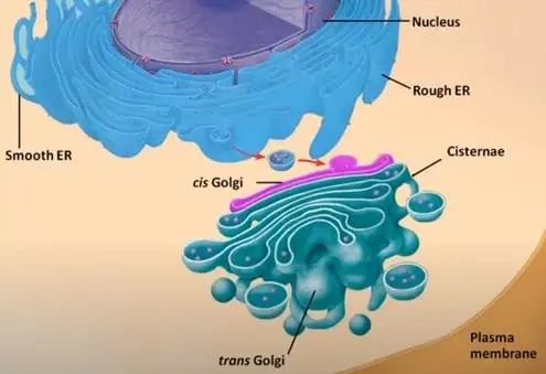
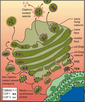
The stacks of the Golgi apparatus are divided into cis-Golgi, the side that receives vesicles from the endoplasmic reticulum and the trans-Golgi, the side that releases the vesicles. The stacks are compartmentalized in the cis Golgi network which has a convex architecture and is closer to the nucleus. The cis, medial and trans cisternae and the trans-Golgi network represent the concave exit face that is closer to the cell membrane and has a complex system of tubules and secretory or coated vesicles (figure 1) (Morré & Mollenhauer 2008, Hawkins 2011, Gartner & Hiatt 2013).
Vesicles are an important part of the Golgi apparatus since they serve as a vehicle to carry proteins from the endoplasmic reticulum, inside the Golgi apparatus and towards its new destination (Morré & Mollenhauer 2008, Gartner & Hiatt 2013). According to Morré & Mollenhauer there are 5 vesicles of the Golgi apparatus:
COP-Coated Transition Vesicles: These vesicles bleb off the rough endoplasmic reticulum and integrate to form new Golgi apparatus cisternae, their membranes are covered by coatomer proteins.
Clathrin-Coated Vesicles: These vesicles can be found at the trans-Golgi face. They are coated with clathrin protein (that have complex spiny coats), associated with functional specialization, transport and fusion.
Secretory Vesicles: These vesicles are formed at the ends of tubules that at the same time are attached to the cisternae. Once the vesicle matures it detaches from the cisternae and migrates to the cell surface to fuse with the plasma membrane.
Secretion Granules: Unlike continuously secreting cells, discontinuously secreting cells accumulate in the cytoplasm as granules. The discharge of these granules requires extracellular calcium and a hormonal or a drug stimulus that promotes secretion.
Fusiform Vesicles and Cisternal Remnants: Fusiform vesicles appear to lack content and only deliver plasma membrane units without any secretion occurring, while cisternal remnants have been suggested to be released into the cytoplasm along with secretory vesicles (Morré & Mollenhauer 2008).
Golgi Apparatus Function
The endoplasmic reticulum synthesizes proteins and lipids that are transported to the Golgi apparatus. Tubules from the Golgi apparatus appear to improve the connection between the smooth membranes of the endoplasmic reticulum and the Golgi apparatus.
While transported, the proteins are modified and sorted to different locations in the cell, such as plasma membrane, endosomes and lysosomes. Golgi enzymes are responsible to support vesicular transport and modifying the vesicle’s contents by glycosylation, phosphorylation, sulfation and proteolytic processing.
The Golgi apparatus is also involved in the synthesis of important molecules such as glycosaminoglycans, phospholipids and sphingolipids (Morré & Mollenhauer 2008, Hawkins 2011).
Golgi Apparatus History
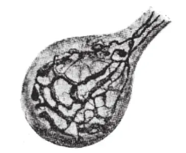
The discovery of the Golgi apparatus was made by Camilo Golgi (Nobel Prize in Medicine along with Santiago Ramón y Cajal in 1906) in 1898 describing an internal reticular apparatus resulted from work based on light microscopy using preparations with silver impregnation techniques developed to visualize neural networks, in these experiments Golgi worked with nerve cells of barn owls and cats (figure 2).
A modification of this method continues today known as the “Golgi -Cox” method that indicates how to prepare nervous tissue for examination under the light microscope (Morré & Mollenhauer 2008).
From 1925 to 1955 there was a controversy about the existence of the Golgi apparatus. We have to remember the difficulties of describing microscopic organelles for researchers at that time and more if you take into account that the Golgi apparatus structure varies from cell to cell, even within the same cell type.
The doubts about the Golgi apparatus existence arise precisely due to limitations in staining techniques that led them to think that the Golgi apparatus was an artifact produced by the precipitation of silver or osmium on the surface, between or inside the vacuoles. In microscopy, an artifact is a structure resulting from sample processing that isn’t an actual feature of the specimen being examined. In consequence, several researchers actually denied the existence of the Golgi apparatus (Morré & Mollenhauer 2008).
Fortunately, the invention of the electron microscope ended the controversy. Dalton and Felix published in 1953 the first micrographs identifying the Golgi apparatus by electron microscopy, this was followed by publications of other authors that led to the general acceptance of the Golgi apparatus as a morphological structure.
As a result of this technological advance, unreliable methods used in histology were abandoned and the 40 years of controversy were erased by 2-4 years of research with the electron microscope (Berger & Roth 1997, Morré & Mollenhauer 2008).
Synthesizing Proteins and Lipids
The function of the Golgi apparatus involves a series of reactions that help synthesize proteins and lipids. One of the main processes of protein synthesis is the glycosylation, where protein processing includes the modification and synthesis of carbohydrate portions of glycoproteins. Glycoproteins are proteins with a glycan linked to it.
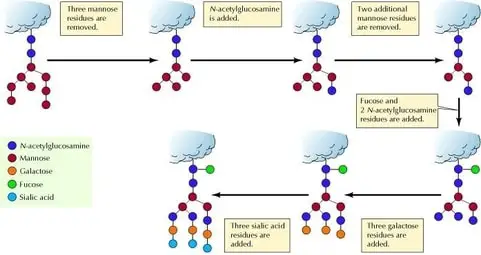
A glycan is a just a carbohydrate that has multiple sugar molecules. Different glycoproteins are modified to different degrees during transportation depending on the structure of the protein and the number of enzymes needed for processing. Processed glycoproteins from the endoplasmic reticulum have the addition of an oligosaccharide, however, the N-linked oligosaccharides of these glycoproteins are extensively modified once in the Golgi apparatus (figure 3) (Cooper 2000).
Oligosaccharides are molecules composed mainly of monosaccharides which are the simplest form of sugar. In addition to glycoprotein processing, the Golgi apparatus also has a role in lipid metabolism, especially the synthesis of glycolipids and sphingomyelin. Ceramide, which is synthesized in the endoplasmic reticulum is converted to glycolipids by the addition of carbohydrates or to sphingomyelin by the transfer of a phosphorylcholine group (Cooper 2000).
Golgi Apparatus and Disease
Alterations in Golgi morphology and distribution in mammalian cells are referred to as Golgi fragmentation. Several factors can cause these morphological changes such as pathological conditions, overexpression of proteins associated with the Golgi apparatus, and pharmacological agents (Fan et al. 2008, Makhoul et al. 2019).
Studies of the Golgi apparatus condition in neurodegenerative diseases have increased in recent years. Neuronal Golgi apparatus has shown morphological alterations in neurodegenerative diseases such as loss of the network configuration (replaced with small and disconnected elements) and reduction in their vesicles and cisternae adjacent to the endoplasmic reticulum. In this sense, the loss of the ribbon in the Golgi apparatus and the aforementioned morphological changes have been related to Alzheimer’s disease, Huntington disease, amyotrophic lateral sclerosis, Parkinson’s disease and other neurodegenerative diseases (Fan et al. 2008, Makhoul et al. 2019).
Golgi fragmentation has been extensively studied in recent times. The precise mechanisms causing Golgi fragmentation are not exactly known. To start understanding the mechanisms by which the changes in the Golgi apparatus affect a person’s health is important to know the different Golgi morphologies during normal processes and under pathological conditions. Despite the fact that technological advances are developing rapidly, many questions remain to be resolved in the research about this organelle (Fan et al. 2008).
Takeaways
To sum up, the Golgi apparatus is a complex organelle with a particular morphology, constituted by a system of stacks of cisternae arranged in a side by side manner with tubules and vesicles.
The functioning of the Golgi apparatus is essential for life to exist since it takes care of protein and lipids synthesis and transportation from the endoplasmic reticulum to specific locations inside the cell or outside the cell.
Unfortunately, alterations in the Golgi apparatus morphology are related to neurodegenerative diseases and their mechanism are not known yet.
The Golgi apparatus may be one of the best and most unique names in cellular biology which is fitting for such a unique and fascinating organelle!
References
- Cooper, G. M. (2000). The Cell: A Molecular Approach. 2nd edition. Sunderland (MA): Sinauer Associates. The Golgi Apparatus.
- Fan, J., Hu, Z., Zeng, L., Lu, W., Tang, X., Zhang, J., & Li, T. (2008). Golgi apparatus and neurodegenerative diseases. International Journal of Developmental Neuroscience, 26(6), 523-534.
- Makhoul, C., Gosavi, P. & Gleeson, P. A. (2019). Golgi dynamics: The morphology of the mammalian Golgi apparatus in health and disease. Frontiers in Cell and Developmental Biology, 7, 112.
- Gartner, L. P., & Hiatt, J. L. (2013). Color atlas and text of histology. Lippincott Williams & Wilkins. 512 pp.
- Hawkins, C. J. (2011). Golgi Apparatus: Structure, Functions and Mechanisms. Cell biology research progress. Nova Science Publishers. 169 pp.
- Morré, J., & Mollenhauer, H. H. (2008). The Golgi apparatus: the first 100 years. Springer Science & Business Media. 307 pp.