

There are plenty of different techniques to use when observing organisms, but the hanging drop slide technique is unique. Other methods work best with viewing a specimen encased in a somewhat solid environment, but this practice is best for viewing live culture with liquid surroundings.
The hanging drop slide technique is used to view live cultures and to inspect the motility or mobility of bacteria. This is accomplished by placing a droplet of fluid in a slide with a dimple in the center, then placing the cover slide on top, which suspends the culture in the concaved area.
The invention of this method revolutionized the microbiology field. It has aided researchers for years and it is still helping in discoveries today.
History of the Technique
Robert Koch was born in Germany in the mid-1800s and began his journey into the field of biology in his early 20s. However, it was not until 1878 that Koch discovered the hanging drop slide technique. He was looking for an easier and faster method of viewing the motility of bacteria. The original way that they had conducted this research before this method took too long and was unnecessarily complex. The hanging drop slide was revolutionary because it solved all these issues in such a simple way.
Today, in addition to using this technique to research motility, researchers utilize this technique to study the presence of flagella in the cells. It is best to use a wet mount like the hanging drop slide to eliminate air bubbles when conducting motility and flagella research. This is much easier to make out the distinct lines, use, and the number of flagella in the sample.
How To Use this Method

It was mentioned earlier that this technique is very different from all others. So it probably goes without saying that it requires tools and steps that may not be used in other techniques. While this creates interesting, new options and experiments, it also comes with the price of having to put in a bit more work to learn how to do this all correctly.
Items needed for a hanging drop slide:
Projects using the hanging drop slide method need specific equipment. To use this method, you must first have the following equipment:
- Cover plate
- Petroleum jelly
- Toothpick or other like tools
- Inoculating loop wand
- Flat plate with a concaved center
Method instructions:
Begin the research by setting down your cover plate and place a small amount of petroleum jelly in all four corners of the glass with a toothpick or something similar with a small, clean end. Next, use the inoculating loop wand to pick the specimen you will be viewing and place it in the center of the cover plate. Once you have secured the liquid on the plate, carefully set the flat plate with a concaved center on the cover plate with the cavity area of the flat plate encasing the sample.
After everything has been safely put together, you must invert the whole thing to allow the droplet to hang in the plate’s dimple and begin viewing the culture with a 10X object lens on your microscope. Then, after you have adjusted the coarse focus and are able to view the cells, switch to the 40X objective lens for a more magnified image. Once you have obtained your notes, clean each of your instruments and go over your research. (Source: Biology Reader)
Why Should You Use this Method?
This technique is often employed when studying the movement and details of bacteria.
“Bacterial motility or mobility can be well studied by employing a hanging drop method, in which a bacterial cell can freely move in the liquid medium.”
Supriya N., a writer for Biology Reader
The liquid aspect allows for easier observation of the typical movements of bacteria and how they are necessary to the life of the organism.
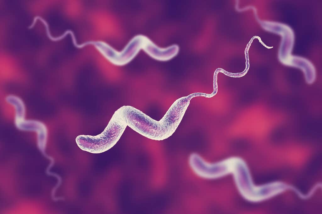
Since the movement is less restricted, the tentacle-like appendage, called a flagellum, can be viewed in a more natural state as well. This is needed to understand the biology of these lifeforms and what their biology is really like.
Why should I research motility?
Motility is the understanding of how an organism is able to move or if it has the ability to move at all. This is necessary to research when figuring out what the bacteria is moving towards or is running away from. If one organism is seen fleeing from another, there is an obvious dynamic between the two organisms that must be observed.
On the other hand, if you are watching a specimen chase another, or move towards something, it can tell you something very different. This all adds up to researching the life aspects of bacteria and how they work with other surrounding organisms.
Why should I research the flagella?
Britannica defines the flagellum as, “Flagellum, plural flagella, hair-like structure that acts primarily as an organelle of locomotion in the cells of many living organisms.” The flagellum characteristic appears in the Mastigophora bacteria, like Chilomonas, Eudorina, Pandorina, and more. It also is present in more common microorganisms like euglena.
Not only does this aspect help define the group the organisms fall under, but also it is necessary to the natural movement. The flagella are used to move around the liquid the bacteria are found in. If you are researching mobility, you must also be looking at the flagella of the entity.
(Source: Britannica)
When This Technique is Not Useful
While this technique is amazingly useful for viewing living organisms, it has its disadvantages as well. When trying to research motile cells, they can sometimes get lost. This means there are so many nonmotile bacteria there, as well that some important organisms can get overlooked when doing research.
To help avoid this, be patient and take your time. Watch carefully for each moving thing and take extensive notes. While setting up this method is quick and easy, the study itself is time-consuming. Just remember to relax and move on only when you are certain that your search is complete.