
There are many different microscopy techniques for one to employ to achieve the desired observation results given the specimen and the specific parts of the specimen that are of interest to the observer. A wet mount slide is a common slide preparation technique used to observe many types of live microorganisms.
A wet mound slide is a slide that is prepared with some type of liquid placed at the center of the slide that will serve as the medium through which the specimen will move or interact with during microscopic observation.
A wet mount slide is by far one of the most common types of microscopy techniques used, so in this post we will discuss why and when wet mound slides should be prepared and the steps to prepare a wet mound slide for yourself.
When to Use a Wet Mount
Wet mount slides are most commonly used where you are observing live specimens that either live in water or their movement is most easily observed in a liquid medium. For example, you could technically observe bacteria under dry mount slide however this will greatly reduce the ability of the bacteria to move. Under a wet mount slide the bacteria can more easily move and their interactions with the environment can be more easily observed.
There are some microorganisms that live in water and require water for survival. In these cases, your only practical option is to use a wet mound slide. An example of this would be paramecium. If you prepared your wet mount slide with a paramecium sample and then let it dry out, you would be left with a dead paramecium and a less than exciting observation.
There are some cases where you are observing a specimen or part of a specimen that is no longer living where a wet mount is not required. For example, if you were observing the leg of a fly there is no need to add a drop of water because there is no movement to be seen and you may actually be contaminating the specimen or adding additional artifacts that you will have to sift through during microscopic observation.
How to Prepare a Wet Mount Slide
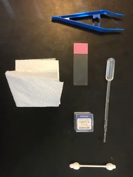
To prepare a wet mount you need a few materials. You can get the majority of these items in this kit.
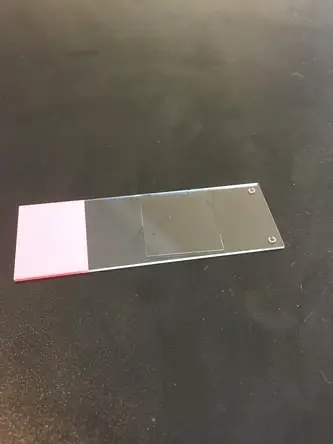
- Standard glass microscope slide
- Pipette
- Specimen sample
- Water
- Slide coverslip
- Tweezers
- Paper towel
- Q-tip
The first step is to use the Q-tip to swab your sample and apply it to the center of the microscope slide. If your specimen is a littler larger you can use tweezers or whatever tool that will help you place the specimen in the center of the slide without damaging it, killing, it or contaminating it. If your sample is some microorganism in the water, you can skip this step.
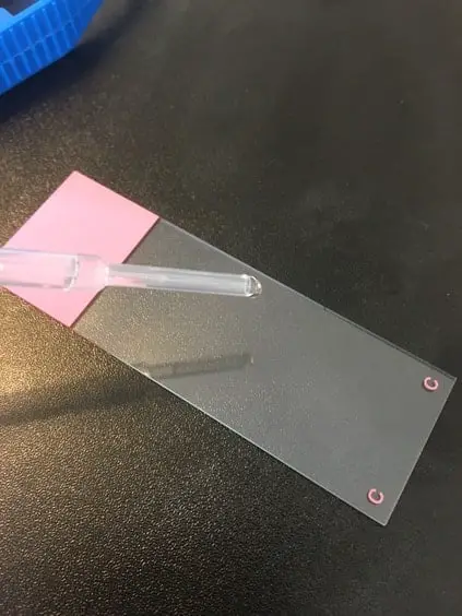
Next use the pipette to place a small water droplet in the center of the slide. This should be a very small droplet. You do not want to get too much water on the slide at a time.
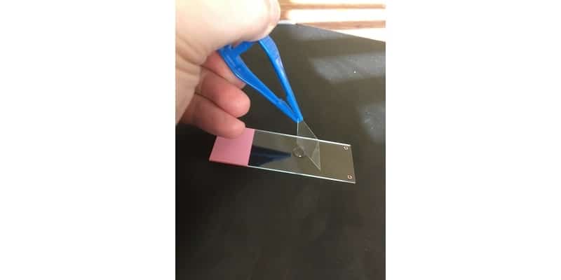
Then, using the tweezers, take the slide coverslip and place one side of the coverslip down on the slide with the other side still above the slide. Use the tweezers to slowly and gently lower the other side of the coverslip onto the slide.
It is important to lower the other side of the coverslip slowly and gently because is you just let the coverslip drop onto the water droplet you will get air bubbles. Air bubbles will make viewing your specimen much more difficult.
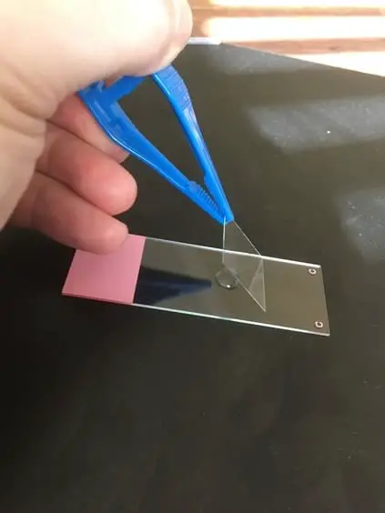
Once the coverslip touches the water it has a tendency to move to make sure as you lower the coverslip you don’t release the coverslip from the tweezers too soon.
If you have added too much water and the coverslip has floated or if water has bled out to the edges of the slide you can use the edge of the paper towel and place it next to the edge of the coverslip and just let the paper towel extract some of the excess water. Now you are ready to take your wet mount slide and place it on the microscope stage and start observing your specimen.
Pro Tip:
If you are observing a specimen that has very rapid movements or is difficult to keep in the field of view because it is constantly moving, you can add a quieting solution that will actually slow the movement of some microorganisms.
This allows you to keep them in the field of view for easier observation and will not inhibit their natural characteristic movements. The most commonly used brand of quieting solution is called Protoslo.
Types of Wet Mounts
Water
The most common type of wet mount is a water based wet mount slide. The reason is that most microorganisms live in some type of water medium and most that do not can still move and survive in it. Water also tends to help preserve organic material and thus is appropriate to be used during long observational periods.
Vaseline
Another type of wet mount is known as the Vaseline Mount. This type of mount is really just used in conjunction with a water wet mount to keep the water from drying out during long periods of observation where the illuminator of the microscope is producing heat that dries out the wet mount. This type of wet mount extends the time that it takes for the wet mount to dry out and preserves specimens that would die as a result.
To prepare a Vaseline mount, use the Q-tip and apply some Vaseline only to the outer edge of the slide coverslip. Then once you have the outer edge coated you can lower the slide coverslip just like you would on a standard wet mount (as described above).
Make sure that you do not touch the center of the slide coverslip with your finger or with the Vaseline or else you may get fingerprints or Vaseline on the coverslip which will distort your image. Next you ensure that you have created a seal that will help prevent evaporation, you can slightly press the edges of the coverslip with a clean Q-tip to ensure a seal is in place.
Saline
Depending on the specimen, water may not be the best medium to view movement for various technical reasons. For example, saline wet mounts are the preferred method to view movements of certain protozoa during the feeding cycle stage. Saline wet mounts are prepared the same way a water wet mount is prepared except instead of water, of course, you use a saline solution.
Common Problems
Air Bubbles
There are some common problems people face during observation when using a wet mount slide. The most common problem is the formation of air bubbles. Air bubbles can form if your slide coverslip closes too rapidly causing air to be trapped under the coverslip or a displacement in the water that is rapidly filled by air.
Air bubbles can even form when you place the slide coverslip on the slide correctly. Sometimes air bubbles are unavoidable. However, the presence of air bubbles does not mean you will not be able to fully observe your specimen. If the air bubbles do not obstruct or impair your observation of the specimen, don’t worry about re-mounting the slide just work around the existing air bubbles.

Floating Coverslip
In some cases, you may find that if you add too much water your coverslip will float after you place it over the water droplet. This can cause you slide coverslip to move to one side or the other of the slide and can also disrupt your sample in some cases. Coverslip floating can be avoided by limiting the amount of water that is dropped onto the slide and by slowly lowering the coverslip on the slide.
Contamination
Contamination can occur if you add contaminated water to your slide that contains other microorganisms or even chemicals that may affect you intended specimen. To avoid contamination, you need to understand your specimen and what things in your water source may cause contamination or unintended consequences.
Takeaways
Understanding how to create a wet mound slide is paramount in microscopy. It is a slide preparation technique that you will use repeatedly, and it is one that does take a bit of practice to get just right. There are different types of wet mounts but the basic steps to preparing them are all the same. I hope this post has given you an understanding of the why wet mounts are used and the steps to try it out for yourself!