
If you’ve ever been swimming in a pond, a lake, or some other stagnant body of fresh water out in nature you may have been wondering what other things were swimming with you. There are the things you may be able to see like fish, turtles, and ducks, but there are so many more things that you can’t see, even if the water is completely clear. That’s because there are tiny microorganisms that live in such bodies of water that you cannot see at all, or at least very well, without the help of a microscope. Chances are one of the tiny microorganisms accompanying you without your knowledge was a species of paramecium.
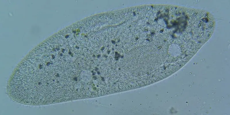
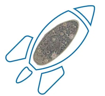
Paramecium is a genus of single-celled, eukaryotic organisms that measure about 50 to 330 micrometers in length across their characteristic footprint shape, which is covered in hair like structures called cilia. Paramecium are found all over the world in freshwater environments and replicate sexually through conjugation and asexually through binary fission.
Paramecium are grouped into a specialized category called “ciliate” because their cells contain small hair like structures on the exterior called cilia which the paramecium use for movement and to engulf their food. Large paramecium can sometimes be visible to the naked eye and will only require a microscope to see the minute details. However, most species of paramecium will require a microscope to see.
Paramecium are officially classified in the following way:
- Kingdom: Protista
- Subkingdom: Protozoa
- Phylum: Ciliophora
- Class: Oligohymenophorea
- Subclass: Hymenostomatia
- Order: Hymenostomatida
- Suborder: Peniculina
- Family: Parameciiade
- Genus: Paramecium
This post is for anyone that wants to get a broad overview of paramecium but also wants to know some of the detailed aspects of this organism. In this post we will look at the anatomy of a paramecium, how it behaves, what is eats, the history, and much more.
Anatomy of the Paramecium
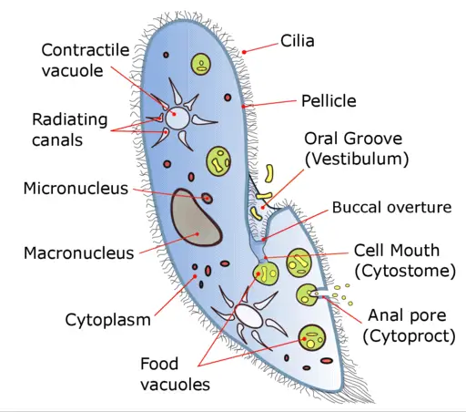
To understand this organism, we need to take a look at what makes this thing tick. Let’s take a look at the anatomy of a paramecia.
Cilia – Cilia are little hair like projections that are just a continuation of the cell surface membrane. The two main functions of the cilia are for movement and for ingesting food. The cilia responsible for ingesting food are located in a funnel shaped depressed region of the cell called the gullet. All other cilia on the paramecium are thought to be used for movement except for the caudal cilia which are longer cilia. During the mating process cilia are used to initiate the mating process also known as conjugation.
Contractile vacuole – There are typically two contractile vacuoles on a paramecium. One located at each end of the cell opposite from the cytostome. The vacuole is used to transport waste liquid out of the cell. The vacuoles work by collapsing in an alternating fashion which empties the liquid out through pores. In short, if there is too much water in the cell, it will rupture, so the contractile vacuole is crucial to the survival of the paramecium. It is constantly working to regulate this balance. There are two different types of contractile vacuoles. One type is a canal-fed vacuole and a vesicle-fed vacuole. The other is called a canal-fed vacuole.
Pellicle – The pellicle is what helps the paramecium keep their shape although it is capable of deformations. The pellicle is made up of three layers; the plasma membrane, the alveolar system, which is a section of flattened membrane bound sacs, and the epiplasm which is layer that lines the inner alveolar membrane. Together these three layers get molded into ridges which actually form shapes like hexagons and parallelograms that appear all over the cell surface.
Radiating canals – Radiating canals absorb wastewater and materials from the surrounding cytoplasm which eventually will get transported out of the cell by the contractile vacuole.
Vestibulum – The vestibulum, also known as the oral groove, is a flattened, funnel-shaped indentation that is the opening to the mouth region of the paramecium. The vestibulum has its own pellicle and cilia. This groove leads into the buccal overture.
Micronucleus – The main purpose of the micronucleus is reproduction. The micronucleus is a generative nucleus that contains the genetic information that is passed along to offspring during reproduction. The micronucleus is located near the macronucleus.
Macronucleus – The macronucleus is ellipsoidal in shape almost like a kidney. The function of the macronucleus controls the metabolism of the cell. The macronucleus lacks a nuclear membrane.
Buccal overture – The buccal overture is an opening that leads to an “S” shaped cylindrical structure called the buccal cavity. The buccal cavity contains four structures called the endoral kinety, dorsal peniculus, ventral peniculus, and the dorsal quadrulus.
Cytostome – The cytostome is the “mouth” of the paramecium and it resembles a tear drop shape. The cytostome transfers the paramecium prey into the food vacuole.
Cytoplasm – The cytoplasm is a jelly like substance that contains the organelles of the paramecium. The cytoplasm suspends the vesicles, ribosomes, and food storage reserves. The cytoplasm also contains everything the organism would need to synthesize proteins.
Food vacuoles – Unlike the contractile vacuole, the food vacuoles do not contract. Food vacuoles accumulate food gathered by paramecium through the cytostome. Then once the food vacuole becomes a certain size it will break off and will travel through the cell. The food will be digested so to speak by enzymes. The useful material will remain in the cytoplasm and the remaining material will be expelled from the cell through the cytoproct.
Cytoproct – The cytoproct, also known as the “anal pore”, is where waste is expelled from the cell. The cytoproct is located, as you might expect, along the rear of the cell.
Trichocyst – It is suggested that trichocysts are used in the defense of the paramecium. The trichocyst has a spindle shaped body and at the wider end and looks similar to a golf tee turned upside down. Trichocysts are located at specialized cortical sites and there are typically about one thousand per cell. When the paramecium is attacked these little filaments are fired at the attacker to try and thwart the attack.
Types of Paramecium
There are 15 different species of paramecium in Aurelia group and a number of other species outside of the Aurelia group. There are other species that have had the classification disputed for several reasons. Paramecium species can be divided into two main groups, primarily by body shape, but also genetically and biochemically.
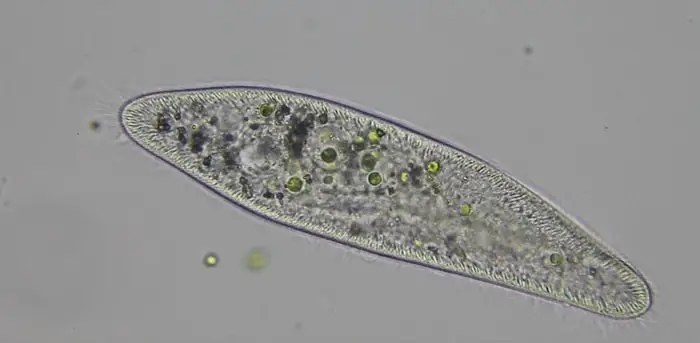
The “Aurelia” group are defined by the relatively long bodies with a pointed end. A few of the common species that fall into this grouping are Paramecium Aurelia, Paramecium Caudatum, Paramecium Multimicronucleatum.
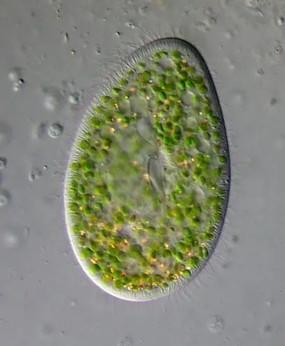
The “Bursaria” group are defined by a shorter and broader body shape and flatter in the dorsoventral position. This group also has a more rounded rear. Some of the species in this group are Paramecium Bursaria, Paramecium Calkinsi, Paramecium Woodruffi, Paramecium Polycaryum, and Paramecium Trichium.
Where do Paramecium Live?
Paramecium are found all over the world. For more than 300 years paramecium have been discovered and observed in many different habitats throughout the world. As long as there is some organic material or decaying matter in a body of freshwater you can bet there is probably paramecium floating around. Fresh water paramecium species can be found in the following places:
- Lakes.
- Ponds.
- Reservoirs.
- Places where there is standing water for long periods of time.
- Aquariums.
- Rivers and streams.
- Pools.
- In some cases, drinking water.
There are other species of paramecium that can be found in the following places:
- Brackish waters.
- Estuaries where rivers or streams meet bodies of salt water.
Although the majority of species are found in freshwater there is one species of paramecium that can live naturally in water that contains a higher salinity than freshwater. Paramecium Calkinsi can live and reproduce in tidal brine pools near the sea.
If you are interested in observing these amazing microorganisms, the microscope I used to capture my photographs and video of paramecium can be found here on Amazon. I have also mounted this DSLR camera to my microscope which has vastly improved the quality of the video and photographs compared the microscope camera that came with my microscope.
What do Paramecium Eat?
Paramecium feed on much smaller organisms than themselves like bacteria, yeast, and algae. Paramecium are able to, in a sense, “smell” or detect bacteria by using indicator chemicals like folic acid which are dissipated metabolites. The paramecium uses these receptors to track down the bacteria.
Once the bacteria are near enough it uses the cilia to push these organisms, along with some water, into the vestibulum. They then move along the buccal cavity until it reaches the mouth (cytostome). From there the bacteria will be acidified and killed.
This will make it easier for the bacteria to be digested by the lysosomal enzymes. From there they get accumulated into food vacuoles which eventually get released into the cytoplasm. After circulating through the cell body, they will be digested by the lysosomal enzymes.
Eventually the vacuoles will shrink when the nutrients all pass into the cytoplasm. After the unused nutrients reach the anal pore they are expelled to the outside environment.
What Eats Paramecium?
Despite the paramecium’s ability to track down and eat its prey, they are not the apex predator in their ecosystem. Some microorganisms that prey on paramecium are amoebas, didiniums, and water fleas. Although paramecium do use trichocysts to defend themselves, they are also able to quickly and effectively rotate 360 degrees to find a means of escape. They can use their cilia to propel themselves quickly away from danger.
Paramecium Behavior and Movement
Paramecium are not known as graceful microorganisms. In fact, if you were to observe paramecium movement under a microscope you would see quick movements in short bursts.
The paramecium moves using its cilia. It propels itself by a coordinated whipping movement by the cilia. Cilia are arranged all around the cell and have a two phase movement. The first is an effective stoke where the cilium is relatively stiff and the recovery stroke where cilium curls loosely and then sweeps forward. These coordinated actions combine to manifest the speedy yet jerky movements of the paramecium.
The paramecium will continue these quick movements until it encounters an object in which case it will quickly move backward to avoid the object. This is known as an avoidance reaction. The paramecium does not have eyes so it will repeat this process until it gets around the object or finds another path.
Reproduction
Paramecium can reproduce sexually and asexually. The paramecium uses transverse binary fission as a means to reproduce asexually. Transverse binary fission which basically means that the paramecium splits perpendicular to the longitudinal axis. Put simply this means it splits in half across the middle as shown in the image below.
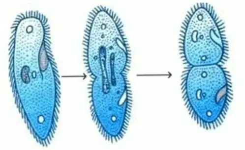
Transverse Binary Fission
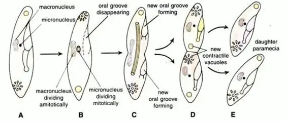
The process of transverse binary fission starts by the division of the nuclei and the disappearance of the oral grooves and the buccal structures. The macronucleus begins dividing amitotically and the micronucleus starts dividing mitotically. What this means in simple terms is that the macronuclei elongates and gets constricted in the middle.
Micronuclei go through the phases of mitosis which consist of the prophase, metaphase, anaphase, and the telophase. Once in the telophase the micronuclei are elongated, two new oral grooves are formed along with new contractile vacuoles.
After the division of the nucleus is complete there is a constriction along the center of the cell which continues to deepen until there is a split and division of the two distinct cells. The two daughter cells are identical to the “parent” because they share the exact same DNA. The process of binary fission takes place about two to three times a day and lasts for about 30 minutes.
Conjugation
The process of sexual reproduction in paramecium also known as conjugation begins with a pair of complementary mating types. The two paramecium come together joining at the cytopharynx region.
These joined paramecium are called conjugates. The region here this union occurs causes the pellicle to disintegrate and then the cytoplasm of each paramecia cell merges together forming a cytoplasmic bridge. Next the macronuclei begin to disappear while the micronuclei begin to divide mitotically as we discussed above.
The micronucleus also divides mitotically to produce four nuclei three of which eventually disintegrate. The remaining micronucleus divides, splitting off into a so called “male” pronucleus and a “female” pronucleus. The two paramecia exchange the male pronuclei through the cytoplasmic bridge and binds with the female nucleus to form a synkaryon also called a zygote nucleus.
Next the paramecia separate, and the nuclei divide through mitosis until there are a total of eight nuclei. Half of the nuclei will start to behave like, and become, macronuclei and the other half will behave like, and become, micronuclei.
Then the two paramecium divide into four daughter cells and then again to form a total of eight daughter cells each with a micronucleus and a macronucleus. In this case there is an exchange of differing genetic material.
How Long can a Paramecium Live?
Although you and I age according to a calendar, it does not seem to work that way for the paramecium. Some studies suggest that instead of aging by calendar days, it may be more accurate to think about the lifespan of the paramecium in terms of number of cell divisions or cell doublings. This is known today as the “Sonneborn limit”.
Sonneborn’s pivotal study used two separate lines of paramecium cells and cultivated them at different temperatures which would induce one group to conduct binary fission faster than the other. This was measured over the same number of calendar days and the resulting data showed a more accurate gauge of lifespan using the number of fissions rather than calendar days.
History
The first accounts of paramecium observation have been credited Antony van Leeuwenhoek as early as 1674. Leeuwenhoek is also credited with building the first “simple microscope”. The handmade microscope used a single lens and used light from the sun or from a candle for illumination.
Fast forward in time a bit and there is some mystery around who might have published the first drawings of the paramecium. In 1703 an anonymous writer wrote a description of and sketched out illustrations of paramecium that was published in the Philosophical Transactions of the Royal society of London. The name “Paramecium” was given to the ciliate group by John Hill in 1752.
The modern classification of the Paramecium Aurelia is credited to Joan Smith-Sonneborn who utilized genetic and biochemical differences along with physical characteristics.
Paramecium in the Classroom
Paramecium are a very common organism to see in a lab for several reasons. Paramecium are readily available in a number of accessible places in the environment, so obtaining a sample is relatively easy.
Paramecium are easily cultivated and are a model organism in certain circumstances to observe primary and secondary endosymbiosis.
Endosymbiosis is where a single celled organism lives within another cell as part of a symbiotic or mutually beneficial relationship. This endosymbiosis is thought to explain or partly explain the emergence of eukaryotic cells from prokaryotic cells in the study of evolutionary biology.
Paramecium and Cigarette Smoke

We all know that cigarette smoke is bad for you, but could it also have damaging effects on paramecium? Even though paramecium live along side millions of microorganisms some of which can cause nasty disease and illnesses in human, paramecium cannot withstand the harmful effects of cigarette smoke.
Researchers in 1967 tested what effect smoke would have on paramecium. The paramecium was exposed to cigarette smoke that was bubbled through distilled water. The paramecium that were exposed exhibited lowered levels of movement and then death only after a few minutes.
Paramecium in Space
You’ve probably heard of monkeys in space but what about paramecium? In this study paramecium were flown into orbit aboard a soviet orbital station where several interesting things were observed.
What they found was that paramecium cultures were proliferating at a higher rate than usual, the paramecium cell volume was larger, there was a difference in cell dry weight, the cell total protein and electrolyte content was also higher.
A possible reason for these observed changes is likely due to the effect of weightlessness on the cell membrane permeability.
Takeaways
Paramecium are incredible microorganisms and have so many things to teach us as a model organism in so many studies and research endeavors. Paramecium are found all over the world and have a multitude of species that vary in size, shape, biochemistry and genetic material.
Paramecium were some of the first microorganisms observed in some of the earliest microscopes in the world and continue to be observed by microscope enthusiasts, biologists, geneticists, and microbiologists alike.
References
- http://kops.uni-konstanz.de/bitstream/handle/123456789/7881/Plattner_My_favorite_2002.pdf?sequence=1
- Wichterman, Ralph. The biology of Paramecium. Springer Science & Business Media, 2012.
- https://en.wikipedia.org/wiki/Paramecium
- Watanabe T. (1990) The Role of Ciliary Surfaces in Mating in Paramecium. In: Bloodgood R.A. (eds) Ciliary and Flagellar Membranes. Springer, Boston, MA
- Allen R.D. (1988) Cytology. In: Görtz HD. (eds) Paramecium. Springer, Berlin, Heidelberg
- https://doi.org/10.1002/jez.1402600111
- https://www.uniprot.org/locations/SL-0268
- https://doi.org/10.1080/00039896.1967.10664819
- https://www.ncbi.nlm.nih.gov/pubmed/1244399
- https://link.springer.com/chapter/10.1007%2F978-3-319-32211-7_16
- https://doi.org/10.1016/0273-1177(81)90249-0
