
In life science, a centriole is a specialized piece of cellular machinery found within most animal and plant cells. While centrioles are crucial for many animal and plant cells to work properly, bacteria and most fungi do not contain centrioles.
The human body contains trillions of centrioles which are the parts that make up a centrosome. Each centriole is approximately 250 nanometers long, or three hundred times the width of a human hair. While they may be small, centrioles are essential cellular components.
Centrioles function to help cells complete their life cycle. The cellular life cycle refers to the sequential series of events a cell completes in order to properly grow and divide. In addition, centrioles function to organize cellular structural components and maintain cellular structure.
Without tight control of a cell’s life cycle and structure, cells will mutate and die. In this context, centrioles play a truly critical and basic role in animal and plant life. With this big picture in mind, it is fitting that the word “centriole” is derived from Latin words meaning “little central part.”
Before going into great detail about what the centriole does, it’s important to understand the centriole’s specially-designed shape, structure, and location within the cell.
Anatomy of a Centriole
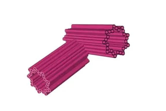
Structurally, centrioles are made of multiple connected pieces of dynamic polymers called microtubules. Microtubules are widespread and indispensable in animal and plant cells. They function to maintain structure within the lively, unorganized fluid of the inner cell cytoplasm. Microtubules make up a structural network within the cytoplasm called the cytoskeleton.
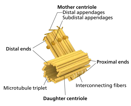
To form a centriole, the microtubules are connected to one another in triplets. These triplets are then connected symmetrically to nine other sets of triplets to form a cylindrical barrel. The centriole cylinder has radial symmetry, and if one was to look down the barrel of the cylinder, the resulting shape is a star with nine points.
Within the cell, centrioles are positioned in the jelly-like cytoplasm that comprises the fluid inner compartment of living cells. In most cells, centrioles are not attached to the outer cell membrane or bound by their own membranes. Instead, they are located in the cytoplasm near the nucleus. In animal and plant cells, the nucleus is a prominent membrane-bound organelle that contains DNA and acts as the cell’s central control center.
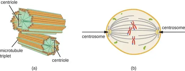
Centrioles located near the nucleus function in complexes made of two centrioles called centrosomes. The two centrioles that make up centrosomes are arranged perpendicularly, at right angles to one another.
Centriole Functions
For the sake of simplicity, this article will focus on presenting an overview of centriole and centrosome function that occurs in most animal cells. Exceptions to this dogma exist in certain plant, yeast, and insect cells. The two predominant functions of centrosomes are to: 1) maintain cellular organization, and 2) maintain proper functioning of the cellular lifecycle.
While centrioles are made up of microtubules, they also regulate the production, location, and organization of other microtubules that do not form centrioles themselves. Instead, these microtubules make up a significant portion of the cytoskeleton.
During microtubule organization, the centrosome, comprised of two perpendicular centrioles, is surrounded by a blob of protein called the pericentriolar material. This material contains proteins that are necessary for the initiation of microtubule organization.
Microtubule organization begins with microtubule nucleation. Nucleation is a process by which single microtubules form into the long filaments that make up the cytoskeleton. This centrosome-mediated organization and localization of microtubules and cytoskeleton structure are very important for proper functioning of the cell cycle. In this way, the two major cellular functions of centrosomes are tightly interconnected.
The second major function of centrioles and the centrosome is to help the cell complete its lifecycle. In order to understand the role that centrioles play in this context, it’s necessary to understand the basic steps of the cellular lifecycle. The cellular lifecycle refers to a series of events that progress in a specific sequence to allow cells to grow, replicate their internal contents, and successfully divide.
When the cell cycle has been properly completed, a progenitor cell will have produced two identical daughter cells. Each daughter cell contains the same DNA and important organelles as the progenitor cell. The part of the cell cycle where centrioles and the centrosome are most important is the organization and partitioning of cellular contents prior to cell division.
The cell division process, called mitosis, starts when cellular components are partitioned into roughly equal compartments that will become the separate daughter cells. When mitosis begins, the cell’s DNA has already been copied and these copies remain inside the nucleus. During mitosis, the centrosome becomes associated with the membrane that surrounds the nucleus.
As cell division progresses, the nuclear membrane breaks down, and centrosome-associated microtubules form a highly structured spindle. This spindle forms a plane of symmetry upon which cell division can occur. The spindle also allows DNA to be partitioned between the two daughter cells. Centrioles are duplicated and partitioned during cell division just like DNA and other organelles.
Following successful cell division, each daughter cell will contain one centrosome. These centrosomes are made up of two perpendicular centrioles, just like the progenitor cell. Research has shown that cell division can occur without the presence of centrioles and the centrosome. However, under these conditions, cell division is very messy.
Without the organizational and structural contributions of the centrosome, mistakes are more likely to occur during cell partitioning and division. These mistakes lead to cellular abnormalities or cell death. Human cells cannot survive without centrioles and the centrosome. Human diseases like microcephaly, dwarfism, and some cancers have been associated with centrosome malfunction. Some tumors contain multiple unnecessary centrosomes.
Evolutionary Considerations
In addition to functioning in cytoskeletal organization and cell division, the location of the centrosome also determines the location of flagella and cilia in cells that contain these motile appendages.
Human sperm cells also rely on centrosomes for cellular propulsion. Evolutionarily, the centrosome and centrioles are ancient structures that evolved over 2 billion years ago. The cilia-containing last common ancestor of all animal and plant cells contained centrioles, underscoring their ancient evolutionary beginnings.
It has been proposed that, over time, the function of centrioles evolved from positioning cilia and flagella to controlling cytoskeleton organization and cell division. There is one major evolutionary mystery regarding centrioles.
Scientists do not fully understand why certain types of plant cells have evolved to no longer contain centrioles. Despite the crucial role of centrioles in biology, described previously in this article, conifers and flowering plants do not contain centrioles.
Evolutionary biologists do not fully understand why this is but speculate that it may have to do with the fact that centrioles serve two major functions (structural organization and cellular life cycle) and one minor function (cilia and flagella positioning) throughout the plant and animal kingdoms.
History
Centrosomes were discovered by Belgian marine biologist Édouard Joseph Louis Marie Van Beneden in 1876. Beneden is the same scientist who discovered chromosomes. While Beneden discovered centrosomes, it was a different biologist, Theodor Boveri, who named them in 1888.
Theodor Boveri was a prominent thinker who contributed to a general theory of chromosomal inheritance in the early twentieth century. The role of centrosomes in microtubule and cytoskeletal organization was discovered first. This remained the sole focus of the field of centrosome research until the mid-1990s, when centrosomes were first associated with cancer.
Then, in 2001, groundbreaking research found that centrosomes were also involved in cellular lifecycle regulation.
New Research
Recently, centriole and centrosome research has been booming, with many new high profile discoveries challenging the existing dogma with regard to centrosome function in cells.
A new study in fruit flies found a site of microtubule organization anchored to the nuclear membrane, and found that it does not require a centrosome. Centrosome research has become a prominent subfield of cancer research in recent years.
Recent cancer research has shown that, not only is overproduction of centrosomes associated with some human cancers, the loss of necessary centrosomes is associated with prostate cancer. In addition to centrosome overabundance and centrosome loss, centrosome size dysregulation occurs during some human cancers. Size dysregulation has been shown to be especially present in breast cancers.
In cancerous cells, overly long centrioles produce overactive centrosomes with overactive microtubule nucleation. The underlying malfunctions in centrosome biology that have been associated with human cancers have also been associated with microcephaly. Microcephaly is a developmental condition where infants are born with smaller than normal sized heads. Known causes of microcephaly are viruses, toxin exposure, and drug use in pregnant women.
Overabundance of centrosomes and loss of centrosomes have been observed in during development resulting in microcephaly. In the past decade, the field of centriole and centrosome research has made major progress in understanding the basic science behind how centrioles and centrosomes are assembled.
In addition to assembly, major progress has been made to understand how these structures function in both disease and non-disease states. Further research in this expanding field will elucidate precisely how centrioles and centrosomes contribute to human cancers, dwarfism, and microcephaly.
Takeaways
Centrioles and centrosomes play extremely important roles in cell functioning and cell division. These tiny gear like structures may contain the key to understanding evolutionary trends at the cellular level and may also be a component in the fight against cancer. I hope this post has turned some of the gears of your mind and will spur excitement for further research!
References
- https://www.sciencedirect.com/topics/neuroscience/microtubule-organizing-center
- https://www.annualreviews.org/doi/full/10.1146/annurev-cellbio-100616-060454
- https://www.ncbi.nlm.nih.gov/pmc/articles/PMC4113101/
- http://nautil.us/blog/the-self_made-beauty-of-the-centriole
- https://www.ncbi.nlm.nih.gov/pubmed/11146294
- https://www.ncbi.nlm.nih.gov/pmc/articles/PMC4113101/
- http://nautil.us/blog/the-self_made-beauty-of-the-centriole
- https://www.ncbi.nlm.nih.gov/pmc/articles/PMC4979663/
- https://link.springer.com/content/pdf/10.1007/s00109-002-0374-y.pdf
- https://www.ncbi.nlm.nih.gov/pubmed/11533726
- https://www.nature.com/articles/s41556-020-0470-7
- https://www.nature.com/articles/s41388-019-0995-z
- https://www.nature.com/articles/s41467-018-03641-x
- https://www.nature.com/articles/nrm.2017.127
- https://www.genome.gov/genetics-glossary/Centriole