
As the most basic of microscopy techniques, bright-field microscopy is also the most common.
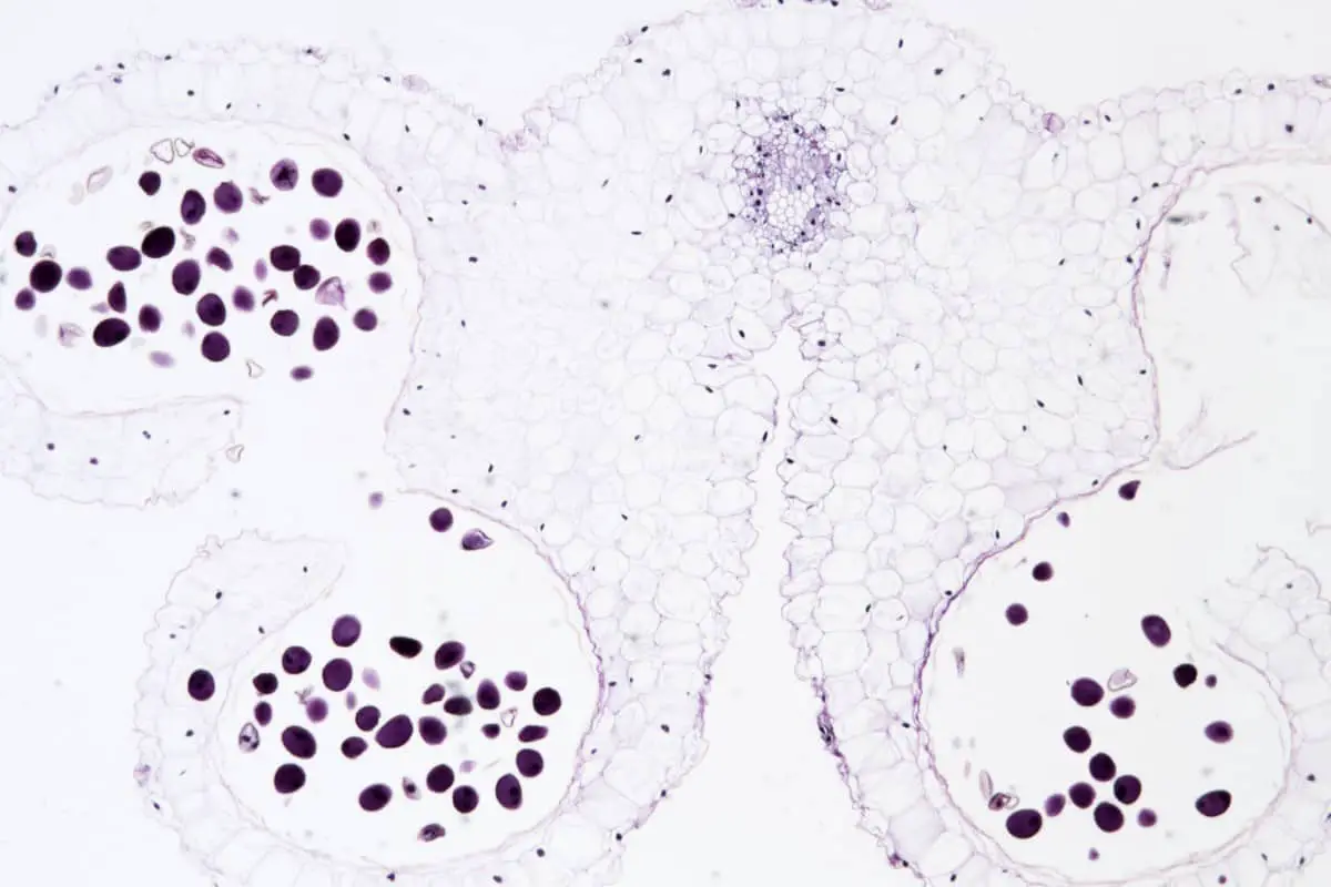
Bright-field microscopy is a very basic, popular technique in which the magnified image appears dark against a white background. Thus, the name: a bright field. It is fairly easy to magnify with bright-field microscopy, but it is a more limited technique compared to others.
In short, bright-field microscopy is most likely the technique your high school science teacher used for their classes. Many modern, easy to access microscopes use this technique, but there’s a lot more to it than you might expect!
Performance
In a bright-field microscope, light travels from the light source, down to the specimen, through the lens and eyepiece, then to the user’s eye. The light is transmitted through the specimen, making it appear dark against an illuminated white background.
Advantages
Because of the simplicity of its setup, a bright-field microscope requires only basic equipment. Nothing beyond the normal light microscope components is required. The microscope is made of these basic parts:
- Eye piece: The eye piece, or ocular lens, creates the final magnification and is used to view the specimen.
- Objective lens: The objective lens collects the light passing through the specimen
- Stage: The stage is where your specimen will be placed
- Condenser lens: The condenser lens collects the light from the light source and directs it to the specimen, which is kept on the stage.
- Light Source: This can be a halogen lamp or an LED light
- Condenser Lens: This collects the light from the light source and directs it to the specimen, which is kept on the stage.
The video below explains microscope mechanisms in more detail.
These are the basics, but the microscope can be adapted with new technology, and optional equipment can be added. Once set up, it requires fewer adjustments to view specimens. Some specimens can even be seen without staining and the optics used in the bright-field technique doesn’t alter the color of specimens. Even living cells can be seen with little difficulty.
Disadvantages
Simplicity makes the bright-field technique popular, but it does have its limitations. The white background has a very low contrast to most biological samples. So, samples that are naturally colorless and transparent cannot be seen well. These samples must be stained, which entails adding a colored dye to the liquid in cells to increase contrast. This is an effective method, but staining may create incorrect details in the specimen.
The second disadvantage is the limit of magnification. The practical limit is around 1300x. Higher magnification is possible, but it becomes difficult to keep the image clear. Because of this, images will be blurry and less detailed.
The bright-field technique requires a strong light source for high magnification. This can produce heat and damage specimens, and can even kill living microorganisms.
Enhancements
Fortunately, many of these limitations can be altered, or fixed entirely, by adjusting the microscope’s equipment. Using sample-staining methods such as simple stains or differential stains, you can add contrast to hard-to-see samples without damaging the specimen. Or, using a colored (usually blue-tinted) lens on the light source can highlight samples that can’t be seen under white light.
An oil-immersion objective lens can decrease the blurring of a sample when magnification is increased. Oil-immersion is a technique in which both the objective lens and the sample specimen are dipped in transparent oil. Immersion oil is like glass—it improves the resolution of the lens and the specimen.
An oil-immersion objective lens can decrease the blurring of a sample when magnification is increased. Oil-immersion is a technique in which both the objective lens and a glass—later placed on top of the sample specimen—are dipped in transparent oil.
By using an iris diaphragm, the light source can be reduced or increased, adjusting the strength of the light. An iris diaphragm is much like the iris in a human eye; it is a thin, opaque circle with an opening (called an aperture), at the center. Its job is to stop light from passing through, except for the light that goes through the aperture. When a diaphragm is placed in the light path of a lens, the aperture regulates the amount of light that passes through.
An iris diaphragm is different from any other type of diaphragm because it is made of “leaves” that can constrict and dilate, just the same as the iris of an eye can grow and shrink to adjust the passage of light.
Brief History of Microscopy
Bright-field microscopy has become a convenient and readily available technology in our world today. However, it has gone through many changes to get to this point.
The idea of magnifying objects with glass lenses has been around since the early 16th century, but it wasn’t until the end of the century that the idea became reality. In 1609, Galileo Galilei converted one of his telescopes into a simple microscope, arguably by accident.
But the credit for the first compound microscope ever created belongs to the father and son spectacle makers, Hans and Zacharias Jannsen. In the very last decade of the century, the two of them positioned two glass lenses one in front of the other, creating a basic microscope capable of adjusting magnification between 3x and 9x.

Robert Hooke made the new science of microscopy more well-known amongst the public, with the help of instrument maker Christopher Cock. Cock made the microscopes, refining the creation process by combining a simple oil lamp with a glass flask filled with water that focuses light. Then he delivered the microscope to Hooke, who published a book about his observations. His drawings and diagrams—magnified to 50x—made the too-small-to-be-seen organisms that he observed more accessible for the wider public.
However, Hooke had difficulty with the magnified image becoming blurry when more than two lenses were used. It wasn’t until the beginning of the 19th century when Joseph von Fraunhofer was able to produce powerful microscopes that kept the magnified image from blurring so much. In the mid 19th century, Ernst Abbe calculated optics for making lenses. Before his work, microscope lenses had been made with trial and error methods; now, they were more uniform. Check out this post if you are interested in learning more about the history of microscopes.