
The human eye has been called a “miracle of design”. Its complex machinery enables us to explore, perceive and understand the world we inhabit. Yet there are molecules that evade the reach of this brilliant organ. Microscopes, with their ability to magnify even the smallest of atoms, have helped humans explore the realm beyond the naked eye. In this article we will look at the evolution and history of the microscope starting from the very first microscope and moving through each technological advancement in microscopes all the way to the advanced modern microscopes of today.
In ancient Greek, ‘mikro’ means ‘of minute size’ and ‘skopion’ refers to ‘means of viewing’. Although the term ‘microscope’ only began gaining popularity in 1625, the journey towards their invention began centuries in advance, in the bellies of the Roman, Greek and Chinese civilizations.
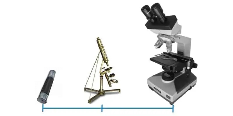
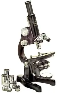
What began as a novelty instrument, accessible only to the elite has now evolved into powerful pen-sized microscopes than can be carried in our pockets. The giant strides humans have made in the understanding of their body, would not have been possible without microscopes. The microscopes available today can be classified in a number of different ways; based on the source of light (light, electron etc), arrangement, number of lenses (simple, compound), or method of interaction between the sample and lens (probe, laser etc). Simple microscopes use the power of a single lens to magnify a given sample. While compound microscopes use an objective lens to collect an image enhanced by a secondary system of lenses.
Based on their differential abilities, each type of microscope has found vital roles in a variety of fields. While some complex electron microscopes assist scientists in visualizing the complex goings-on of cells, others have found use with hobbyists interested in exploring the unseen world around them. With this article, we aim to trace the evolution of this indispensable device; all the way from the birth of the field of optics to the invention of ultra-sensitive digital microscopes.
Optics: The Beginning
The history of microscopy cannot be told without beginning with the field of optics. Glass pieces or lenses made from ground glass have been excavated from many ancient civilizations. The most popular discovery was the Nimrud lens dating back to the Assyrian civilization (modern-day Iraq), 710 BC.
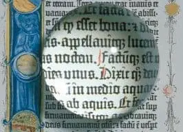
The phenomenon of glass-magnification was known to both the Greeks and Romans. However, the year of this discovery remains unknown. Most experts attribute the finding to mere chance; maybe one day, someone noticed that objects seen through a bowl filled with water appear larger than they are. Euclid (300 BC) was the first philosopher to record this effect. In his magnum treatise ‘Optica’, Euclid writes that the text seen through a glass ball filled with water appeared larger.
Seneca (4 BC – AD 65), a Greek philosopher expanded on this theory, observing that the degree or scale of magnification depended on the angle between the eye and the glass. He astutely associated magnification with the bending of light rays as it travels from one medium to another.
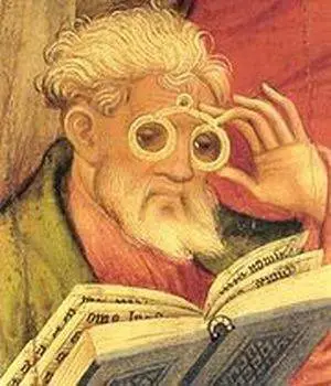
Observations on optics and magnification are littered throughout Greek and Roman records, however, it was not until 1000 AD that lenses called ‘reading stones’ began to be widely used as reading aids to magnify text. The Greeks also used curved lenses made by grinding glass to start fires! Not in the literal sense, but the heat generated by focusing light was used to cauterize wounds, lesions and cuts made in surgery. However, it would be another 1200 years before these curved glasses would find use in spectacles.
Concurrently, several scholars in the Arab world were also working toward understanding optics. Ibn Al-Haytham published ‘The book of optics’ in 1021 AD, summarizing his findings on light and vision. At the time it was thought that light reflecting from the eye ‘touched’ objects, thus enabling us to see them.
There is also evidence to suggest that Chinese civilizations understood and used magnification over 4000 years ago. The Chow-Foo dynasty developed instruments we now call ‘water microscopes’ which consisted of a long tube filled with varying levels of water depending on the magnification required. These simple but effective instruments were capable of achieving a magnification over 150-fold.
In the western world, inventors began experimenting with lenses only in the 13th century. In 1284 AD, Italy, Salvino d’Aramento Degli Amati is thought to have invented the first wearable spectacle. By the 14th century, these eye glasses came to be widely used across Europe. 300 years later, the Renaissance had arrived, aiding scientists from all over Europe to communicate freely. Thus, ushering in the invention of microscopes.
The Early Microscopes
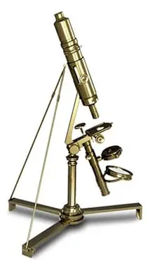
Galileo Galilei (1564 AD – 1642 AD) is widely credited with inventing the telescope capable of magnifying objects at a distance. He then quickly discovered that rearranging the lenses in the telescope with a shorter distance between them helped magnify little things, thereby building the first compound microscope he called ‘Occhiolino’. The device achieved magnification several fold higher than widely used magnifying glasses.
At the same time, Dutch spectacle makers, Zaccharias Janssen and Hans Lipperhey also claimed to have independently built a compound microscope using a combination of tubes and two lenses; the objective lens located over the specimen and an eye piece to view the image. Although it is unclear who first came up with the design for a microscope, the term was widely being used across Europe by 1625 AD.
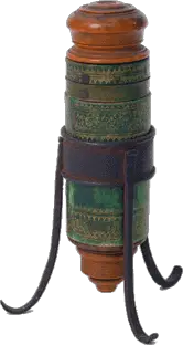
In the same century, Anton Van Leeuwenhoek built the first simple microscope, using a very small but strong lens. He had experimented extensively with grinding and polishing glass, developing curved glasses capable of significant magnification.
Interestingly, it took 150 years to build compound microscopes that could replicate the magnification of Leeuwenhoek’s simple home-made microscopes! In 1674, he used his device to observe bacteria in a drop of water, earning himself the title ‘Father of Microscopes’.
An English physicist, Robert Hooke, Leeuwenhoek’s contemporary was also using microscopes to investigate biological organisms. He used a simple microscope illuminated with candle light to discover cells and their characteristic honeycomb structure. In 1665 the term ‘cells’ was coined and published in his seminary work titled ‘Micrographia’ known for its vivid illustrations of skin and hair. Interestingly it would take over 200 years for scientists to agree cells were the basic units of life.
However, the early microscopes were hard to use and restricted by the quality of their lenses. Over 200 years later, in 1729, Chester Moore Hall developed the achromatic lens. An achromatic lens combines two lenses of different shapes and focal lengths in order to provide better focus.
The Nineteenth Century
The end of the 18th century and beginning of the 19th century saw many advancements in the design and arrangement of lenses thus vastly improving the quality of microscopes. The devices got considerably smaller while improving in magnification.
Additionally the development of new lenses solved common optical problems often seen in the older devices. At this point in time, the journey of evolution of microscopes spreads around the world, with inventors from different countries working together to build better devices.
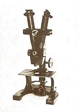
In 1830, Joseph Jackson Lister made massive strides in correcting a phenomenon called spherical aberration. Up until then the image viewed through a microscope would distort based on the angle between the lenses.
However, Lister built a device by placing weak lenses at precise distances from each other which eliminated spherical aberration. This tremendously aided the naturalists of the day in observing distinct biological structures which up until then were just blurred distortions.
The American, John Leonard Riddel was a man of many hats; a scientist, chemist, botanist, geologist, doctor, microscopist, politician and author. Working at Tulane university, in 1850, he built the first binocular microscope with two working eye pieces. Using the device he also pioneered the investigation of Vibrio Cholerae, the bacteria that causes Cholera.
A decade later, the microscope manufacturing company, ‘Ernst Leitz’ built microscopes that incorporated five lenses of different magnifications which could be adjusted based on requirement. They achieved this by fixing multiple lenses on a movable turret located at the end of the lens tube. Concurrently, a rival company ‘Zeiss Optical Works’ was also working toward advancing microscopy.
In 1866, Ernst Abbe while working at Zeiss laid down the principles that guides computational optics development to this day. With the advancement of physics, microscope designs came to favor proved theories over trial and error methods. Using the newly discovered wave properties of light, Abbe built over 17 objectives, each with different magnifications. He also developed the first immersion objectives, in which both the sample and the lens is immersed in a liquid that has a higher refractive index than air.
He developed the mathematical formula called the ‘Abbe Equation’ that helps calculate the maximum resolution of a microscope. But perhaps his best known contribution to microscopy is the invention of the Abbe Condenser, a device placed over the light source in the microscope that concentrates light into a single cone.

At the end of the nineteenth century, many manufacturing companies began designing and building precision microscopes on a large scale. The device became accessible to a wide range of audiences as the prices began going down. High precision microscopes aided some of the most significant achievements in biology including the discovery of chromosomes by Walter Flemming in 1879.
In 1893, August Kohler working at Zeiss Optical works built the patented Kohler illumination device. Up until then condensers were limited in their ability to focus light on the specimen. Kohler’s device vastly improved the resolution and contrast of microscopes by providing an even source of light.
Modern Microscopy: 1900’s – Now
Microscopes enabled scientists and scholars to explore the unseen world around them. The 20th century saw many deadly diseases being understood and cured aided primarily by the visualizing capabilities of the microscope. This century also saw the development of a mass market for microscopes with the company Leitz claiming to have sold over 50,000 devices in the US. Advancements in sample illumination and sources of light enabled the design of novel microscopes which are briefly explained below.
The Ultra Microscope
In 1903, Richard Zsigmondy working at Zeiss Optical Works (Now called Carl Zeiss AG) built the ultra-microscope that enabled the use of waves below the wavelength of visible light (e.g. UV Light). This is achieved by using a light source that scatters light as opposed to older microscopes that simply reflected light.
The device enabled scientists to view particles as small as 4 nanometers by immersing them in fluid. Ultra-microscopes have enabled discoveries in the study of colloids, aerosols, ions and biological ultra-structures. In 1925, Zsigmondy was awarded the Nobel Prize in Chemistry for his discovery.
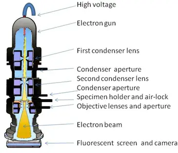
Electron Microscopy
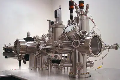
The early 20th century saw the development of a range of illumination sources alternative to light. Electron microscopy uses a beam of electrons to generate an image. The first transmission electron microscope (TEM) was developed in the 1930’s by two German physicists working for Siemens, Max Knoll and Ernst Ruska.
Light microscopes are naturally limited by the physics of light, with a theoretical magnification limit of 500x or 1000x and a resolution limit of 0.2 μM. Using electron beams allows for much higher resolutions, enabling resolution at the nanometre scale. The interaction between the electron beam and the sample is recorded and converted into an image. Advancements in semiconductors and nanotechnology which gave rise to all the advanced technologies in use today was enabled by the TEM. The TEM also enabled the discovery and identification of viruses responsible for many deadly diseases. The TEMs in use today are capable of resolving objects as small as an atom.
In 1942, Ernst August Ruska improved upon the design of a TEM to build the first scanning electron microscope (SEM). The device uses a beam of electrons moving across the surface of a specimen which is then collected to produce a ‘backscatter’ pattern. Although these devices are less powerful than the TEM, they produce high resolution, distinct, sharp, three-dimensional images.
SEMs have found use in biology, chemistry and metallurgy in elucidating the morphology, composition and topology of samples. Ernst Ruska was awarded the Nobel Prize in Physics in 1986 for his design of the electron microscope. Ruska’s principle devices continue to be used till date with magnification capabilities of over 2 million fold.
For more on electron microscopy see Electron Microscopes Explained: From Physics to Images.
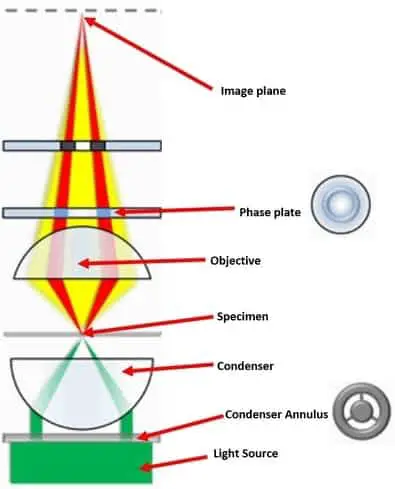
Phase Contrast Microscope
In 1932, Frits Zernike designed a phase contrast microscope which enabled the magnification of transparent samples. The device uses the principle of light interference as opposed to traditional microscopes that are based on light absorption. When light interacts with a medium, the phase or amplitude of the ray changes. However, these phase changes are invisible to the naked eye.
Phase contrast microscopy converts the changes in phases to changes in brightness. The device has been particularly significant to the field of biology. Up until this point, cells had to be mounted and stained, essentially killing them in order to be magnified. However, the Phase contrast microscope enable the imaging of live cells and organelles in their natural state. In 1953, Zernike was awarded the Nobel Prize in Physics for his invention.
Differential Interference Contrast Microscopy
The principle behind differential interference contrast (DIC) microscopy is complimentary to phase contrast microscopy. In contrast to phase contrast, these devices convert the changes in the path of light travelling through a specimen into brightness, with the image appearing as a gradient of black to white against a grey background.
The effect is called Nomarski interference contrast, named after its founder, polish physicist Georges Nomarski. DIC also allows for the imaging of transparent biological samples without staining.
Scanning Tunneling / Scanning Probe Microscope
In 1981, Gerd Binning and Heinrich Rohrer working at IBM, Switzerland built the first scanning tunneling microscope based on their findings of the quantum tunneling (QT) phenomenon. The QT phenomena observed that a small flow of electrons or a small current occurred between a sample and a probe. Scanning probe microscopy measures this current while a small delicate probe moves across the surface of the sample.
With the elimination of the light source, the STM overcomes the shortcomings of both light and electron microscopy. Interestingly, Binning and Rohrer also built in a feedback loop into the STM which regularly adjusted the distance between the sample and the probe. The STM was therefore able to image layers of atoms within the sample, enabling three-dimensional magnification. STMs have contributed to significant findings in both academic and industrial sciences. Today these STMs have ultra-fine piezoelectric probes that are capable of atomic resolution. Binning and Rohrer shared a Nobel Prize with Ernst Ruska for their ground-breaking invention.
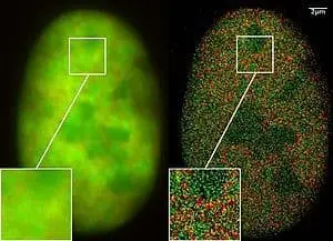
Fluorescence Microscopy
Some substances are capable of absorbing parts of light that is shone upon them to emit light of a different color (wavelength). This phenomenon is termed Fluorescence. For example, the commonly used molecule Fluorescein absorbs blue colored light (high energy) to emit green colored light (low energy). Fluorescence microscopy images the sample by using chemical staining of both live and fixed cellular structures with fluorescent dyes.
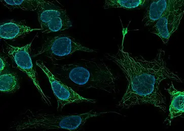
In 1962 the Green fluorescent protein (GFP) was first discovered in jellyfish by Osamu Shimomura, Frank Johnson and Yo saiga for which they shared the Nobel Prize in Chemistry.
This discovery combined with the cloning of GFP in 1992 enabled mass production and wide spread use of GFP in Fluorescent microscopy. GFP based microscopy has fuelled many significant developments in biology, such as elucidation of nerve development, brain development and cancer growth.
Fluorescent microscopy enabled the understanding of critical illnesses including Diabetes and Alzheimer’s. More recently, advancements in gene editing have enabled live cells to express their own GFP without any additional staining methods.
Confocal Microscopy
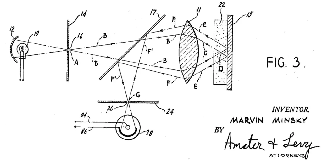
In 1957, Marvin Minsky first proposed confocal imaging as an alternative to light microscopy. In this phenomenon, light is focused on a single point in the sample as opposed to illuminating the entire sample. However, it was not until 1978 that the discovery of lasers aided the design of high-resolution confocal laser scanning microscopes.
Thomas and Christoph Cremer developed an investigating probe with a miniature optical system that investigates light scattering at a precise point in the sample. Working across the sample, the device builds a three-dimensional image of resolution between 0.5–3.0 μm. With the discovery of fluorescent dyes, specific cell structures could be stained, allowing confocal scanning probes to identify their morphology.
Today, confocal endomicroscopes have been developed that do not require tissues to be removed from the body. The device can provide instant histopathology of biological tissue inside the body. This has proven particularly useful in imaging the gastrointestinal tract and its disorders.
X-Ray Microscopes
These devices use electromagnetic radiation, usually X-rays to build three-dimensional images of objects. The most commonly used X- ray microscope is the CT scanner (computational tomography) which allows for the non-invasive imaging of human tissue.
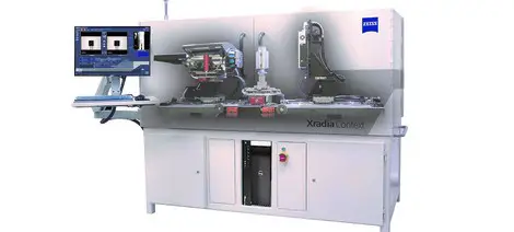
X-Ray absorption patterns across the sample are collected by a computer and used to build a three-dimensional image. Since various tissues in the body absorb X-rays differently, the device can be used to image and observe specific biological structures.
In 1972 the first CAT scanner was built by Allan Cormack and Godfrey Hounsfield for which they were awarded the Nobel Prize in physiology and medicine. Most commonly used in medicine, the device has also found use in non-destructive material testing in industries and archaeology.
Super Resolution Microscopy
The theoretical limit for resolution in a microscope is limited by the diffraction of light, as mathematically defined by Ernst Abbe in 1873. However, the super resolution microscope pioneered by Stefan Hell in 1996 overcomes this limit by combining several different optical techniques.
The devices grouped under super resolution includes the use fluorescent microscopy, photon tunnelling microscopy, super resolution optical fluctuation imaging (SOFI), stimulated emission depletion microscopy etc.
Eric Betzig, Stefan Hell and William Moerner shared the Chemistry Nobel Prize in 2014 for their combined advancement of super resolution microscopy. Today these devices are capable of visualising particles smaller than 0.2 micrometres.
Cryo-electron Microscope
The device is a modification of transmission electron microscopes (TEM). While TEMs use a beam of electrons to examine samples, most biological materials degrade in these conditions.
Jacques Dubochet, Joachim Frank and Richard Henderson developed a novel cryo-electron microscope which allowed for the high resolution of biomolecules by freezing them.
The device also uses a gentler electron beam which does not affect biological structures thus enabling the visualization of proteins, DNA and other bio-molecules as they move and perform function. In 2010, these devices enabled the visualization of atoms in a virus. The 2017 Nobel Prize in chemistry was awarded to these scientists who pioneered this device.
Other Types of Microscopes
A wide range of microscopes are currently in use around the world. Acoustic microscopes use sound waves as opposed to light to examine samples. This allows for samples to be imaged non-invasively. The most common modification of acoustic microscopes is the ultra-sound.
Digital microscopes developed in 1986, uses a digital camera and a computer to carry out live-imaging. Some devices are equipped with eye pieces while others are completely controlled by a computer. The computer is able to analyse properties of the image that escapes the naked eye, including measurement of distances, strength of fluorescence, minute changes in thickness etc.
Dino-Lite digital microscopes are a recent innovation which has found popularity among hobbyists. These are handheld devices, smaller than a pen that are capable of up to 500X magnification.
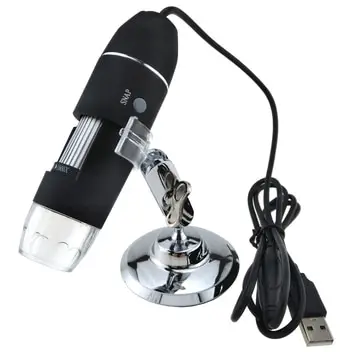
The USB computer microscope is a low-powered digital microscope where the mounted digital computer can be directly linked to the USB port of a computer. Although they are only capable of magnification up to 200X, they are popular for their ease of use. Pocket digital microscopes are particularly suited for children and amateur hobbyists for hand held imaging of low magnification (25X-100X).
Stereo-dissecting microscopes allow for the stereoscopic imaging of samples. The device combines two pairs of distinct eyepieces and objectives to elucidate the depth of samples by producing an erect three-dimensional perspective.
Other microscopes currently in use include comparison microscopes, inverted microscopes, fiber optic inspection microscope, petrographic and polarising microscopes, Two-photon microscopes and Tip-enhanced Raman microscopes.
Takeaways
We’ve come along in the evolution of the microscope from the very first microscope to some of the most advanced microscopic systems in history. They are not only highly technically advanced, but as the cost of these devices goes down, they are becoming highly accessible to armatures and hobbyist that have a passion for science and microscopy.
This is creating interest among the younger generation and creating the scientists of the future that will make the next advancements, that we will some day marvel at, in the evolution of the microscope.
References
- J van Zuylen, The microscopes of Antoni van Leeuwenhoek. https://doi.org/10.1111/j.1365-2818.1981.tb01227.x
- David Bardell, The invention of the microscope. https://doi.org/10.1893/0005-3155(2004)75%3C78:TIOTM%3E2.0.CO;2
- Douglas B. Murphy and Michael W. Davidson. Fundamentals of Light Microscopy and Electronic Imaging
- William Rosenthal, Spectacles and Other Vision Aids: A History and Guide to Collecting
- Lauren Cox, Who invented the microscope. https://www.livescience.com/39649-who-invented-the-microscope.html
- Clara Sue Ball, The early history of the compound microscope. https://www.jstor.org/stable/4606667?seq=1
- Sakurai, T. Advances in Scanning probe microscopy
- Erko,A. Modern developments in X-ray and neutron optics.
- Liz Logan. Early Microscopes Revealed a New World of Tiny Living Things
- Martin et al., The Scanning Electron Microscope. https://journals.sagepub.com/doi/pdf/10.1177/21.2.109
- Kuo-Hsing, Optical Versus Virtual Microscope for Medical Education: A Systematic Review. https://doi.org/10.1002/ase.1844