
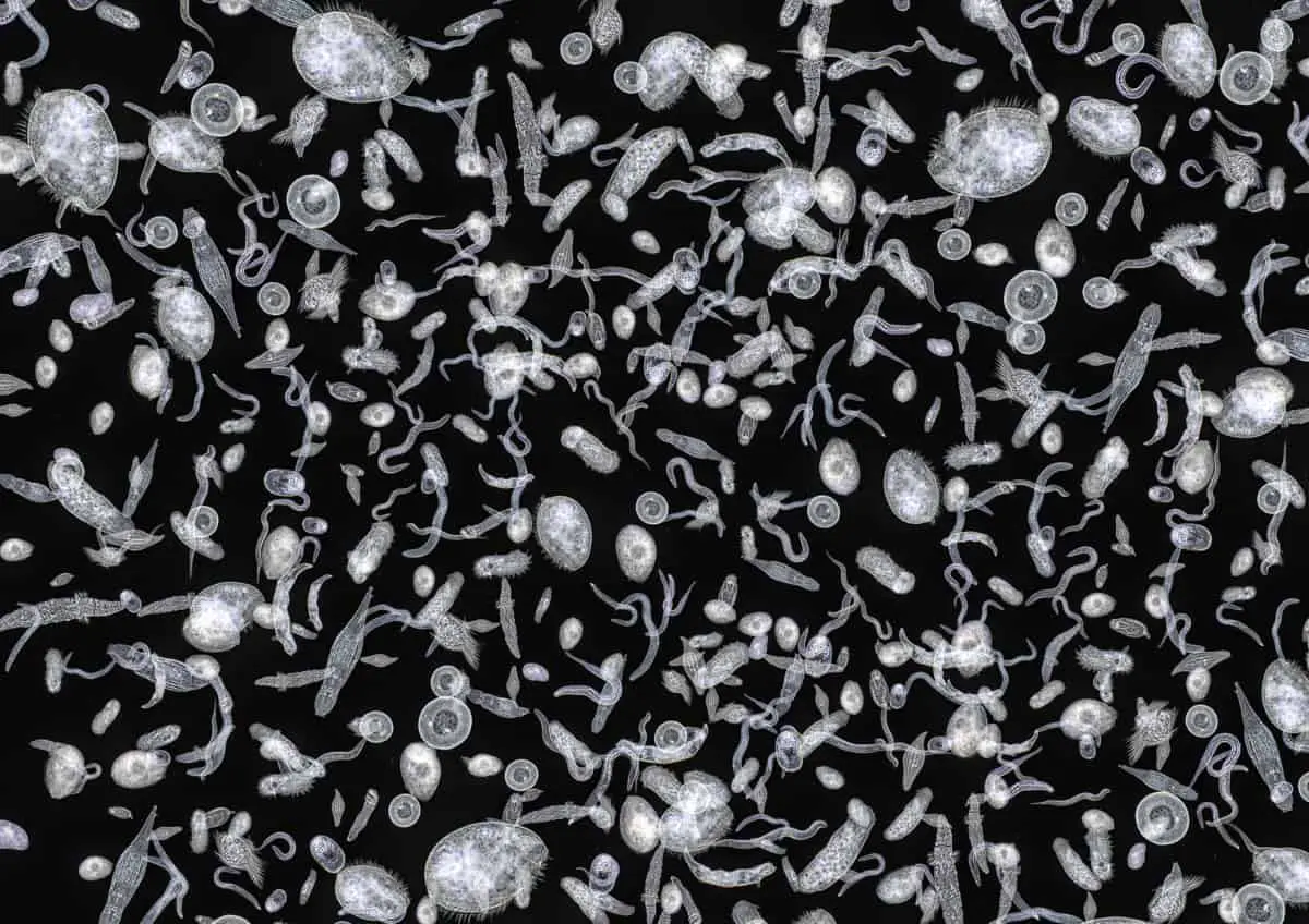
When you look up at the nighttime sky and see stars, those shining celestial bodies are in contrast against the dark backdrop of the nighttime sky though they are billions of light-years away. Dark field microscopy functions very much the same way with small, nearly invisible objects appearing on a dark background to produce stunningly beautiful images.
Dark field Microscopy, also known as dark field illumination, is a method that uses microscopes to make a specimen appear bright white. This effect is achieved by removing dispersed light so that the specimen is appearing through scattered light only. Doing this produces a bright white image on a dark background.
In this article, we will discuss the principles of dark field microscopy. This includes what it is, how it works, what it is used for, and some of its advantages and disadvantages.
Dark Field Microscopy and How It Works

The dark field of microscopy is the principle of making transparent microscopic objects visible. This is done by placing an opaque disc underneath the condenser lenses of a microscope so that only scattered light reaches the eye. The effect of this yields better resolution as objects in the image produced by the microscope will appear visible in bright white illumination against a dark black background.
The condenser lenses of the microscope focus the light towards the sample being examined. By using a dark field condenser, the light is scattered in all directions. The dispersed light is then blocked out by the condenser, allowing only the scattered beams of light to reflect back to the eye. This makes the examined specimen appear on a dark background.
Using this method requires you to have dark field condensers. These are the condensers that refract the non-direct light. Therefore, it is important to make sure the numerical aperture of the dark field condenser is larger than the objective lenses you use. Failure to do so will allow direct light to enter your objective lenses, which disrupts the dark field effect.
There are two types of dark field condensers: dry and immersion. Dry can focus on specimens that require less magnification, producing a dark field image on lenses with a numerical aperture of .65 or less. For specimens that require greater magnification, an oil immersion condenser is needed to produce a high-quality dark background image.
What is Dark Field Microscopy Used For?
This method is great for viewing things that are unstained, transparent, and absorb very little light. Therefore, this method illuminates objects that would otherwise be very hard to see. It also best for examining specimens at low magnification. This is a great method to use for liquid samples, even those with debris in them.
A great example for a liquid sample would be to examine a sample of pond water to see if it is contaminated or to simply observe every living organism contained in it. A sample of this nature is best to be examined through dark field microscopy. Here are a few other examples of what dark field microscopy is used for:
- Examining thin bacteria. Bacteria under normal conditions of microscopy will not all appear in the objective lenses since some appear larger. With the dark field condenser, they will be more visible.
- This method is great for examining tiny marine organisms including algae, bacteria, and plankton.
- Even microscopic dust particles can be examined on the dark background, producing stunning illuminated images.
Basically, the tinier the sample, the better it is to use dark field illumination. This is similar to stars. Stars are billions of light-years away and appear to be relatively small. Yet on the dark background of the night sky, we can see them. The same is true in dark field microscopy; small objects are best viewed on a dark background.

Advantages of Dark Field Microscopy
Observing liquid samples and microscopic organisms that live in aquatic ecosystems is what dark field illumination is best used for. It is always reliable when it comes to these kinds of specimens, and you cannot go wrong.
Examining mounted cells and tissues under a dark field microscope is also very useful.
Objects also appear white on a dark background, which will show fewer details in the specimen. This could be an advantage if you are looking to focus on the external details of specimens.
Lastly, there is an increasing employment rate in its field. If you are interested in working in a scientific field that requires you to use a microscope, dark field microscopy seems to be gaining in popularity due to its combination with fluorescence microscopy.
Disadvantages of Dark Field Microscopy
Images can sometimes be inaccurate or distorted. This is often caused by improper preparation of the slides and their quality. Cleaning the slides is very important as specks of dirt will show up on the sample while you examine it and can degrade your image.
Specimens need to be extremely thin or else the image quality will be reduced. Thin specimens reduce the artifacts resulting from the diffraction process.
Using water and oil on the condenser can result in air bubbles. These bubbles can lead to inaccurate and distorted images.
This method also requires a lot of light to produce the best quality images. This is because the method relies heavily on scattered light to illuminate objects that already absorb very little light themselves. Unless there is a good quality light source, it will be hard to produce the best quality image.
Takeaways
Overall, it is worth the investment to use dark field microscopy. This is mainly because it produces wonderful and beautiful images that could not be obtained in any other way. It is a unique method and is gaining popularity for this reason along with the combination of it with fluorescence microscopy.
Dark field microscopy is best used for illuminating thin specimen samples. The image produced will magnify the tiniest of particles which will appear white on a dark background.