
An Inverted microscope is a microscope where the objective lenses are mounted below the stage and collect light that travels downward through the specimen to the objectives lenses below to form the magnified image. Inverted microscopes do not require specimens to be “fixed” on a slide, thus enabling the magnification of whole organisms, large metal samples and live cells in culture mediums. Even large, heavy industrial samples can be magnified by these devices.
Traditional compound microscopes serve many purposes in both life science and industrial research, but these devices are limited by the size of the samples they can accommodate. In an upright microscope, thin slices of the specimen must fit in-between the objective lens and the mounting stage. This limitation was overcome with the invention of the inverted microscope where the objective lens is placed below the mounting stage.
History of the Inverted Microscope
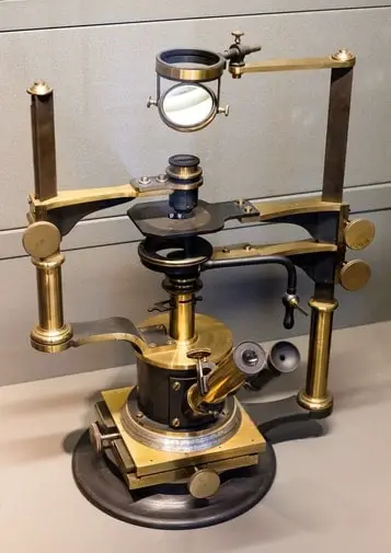
Since the Dutch optician, Zacharias Janssen built the first simple microscope in 1590, many inventors have continued to modify the device to improve magnification and resolution. In 1850, John Smith flipped the traditional design to build an inverted microscope. With this device, large samples could be magnified without cutting them into thin slices.
Before the Second World War, these devices primarily found use in the metal industry, investigating metal samples for composition, coatings, grain size, and defects. In the second half of the 20th century, the study of life once again became the principal focus of scientists.
With the inverted microscope, biological cells did not have to be killed and stained in order to be magnified. This allowed for landmark discoveries in biology, including the discovery of Mycobacterium tuberculosis and cell division.
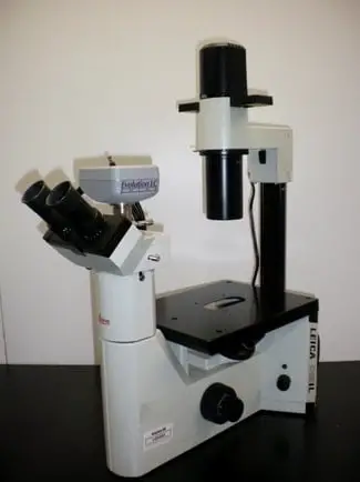
Parts of an Inverted Microscope
An inverted microscope is built with much of the same parts as the traditional compound microscope. However, the parts are simply arranged differently to change the path of light. Light from the light source moves down through the condenser and refracts through the specimen placed on the mounting stage.
A photographic viewing device is placed below the objective to catch the reflected light and form a magnified image. However, this simple design can be modified in several different ways for fluorescence, phase contrast, confocal or digital microscopes.
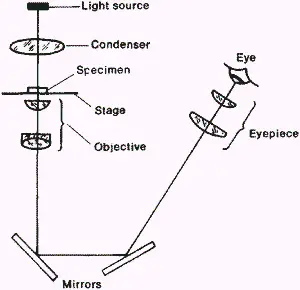
- ILLUMINATION SOURCE: Traditionally, the illumination source consists of a simple halogen bulb housed inside a light chamber, fitted with a diaphragm and condenser. A diaphragm simply adjusts the amount of light that is emitted from the source, thus allowing for varying brightness levels. In modified inverted microscopes such as fluorescence microscopes, halogen lamps are replaced with monochromatic sources such as xenon lamps and lasers.
- CONDENSER: The condenser fitted right below the light source concentrates the light into a single cone of light. The concentration of light helps produce a much sharper image.
- MOUNTING STAGE: The mounting stage in an inverted microscope is generally fixed and cannot be moved.
- OBJECTIVE LENS: Light refracted through the sample is captured by the objective lens to form a real image.A set of objective lenses are mounted around a nose piece to allow different levels of magnification of the specimen. The focal length i.e. the length between the specimen and the objective can be adjusted using the coarse and fine focus knobs. For more on the coarse and fine focus knobs see Microscope Coarse Adjustment and Fine Adjustment: Explained.
- EYEPIECE/ PHOTOGRAPHING DEVICE: A simple set of eyepiece lenses or a complex photographic device can be used toconvert the real image produced by the objective lens into a virtual image that can be seen with the eye.
Why Use and Inverted Microscope?
The inverted microscope allows for significant advantages over the traditional compound microscope. Working distance is defined as the maximum distance allowed between the mounting stage and the objective lens to achieve sharp resolved images. In an inverted microscope, the objective lens is placed below the specimen stage, and the objective itself can be moved to adjust the focal length. Thus large samples can be magnified without affecting the resolution of the image. A few more advantages of the device are summarised below:
- Versatility: Inverted microscopes allow samples of varying sizes and weight to be magnified. Currently used devices can accommodate over 30kgs thus making them ideal for industry use.
- No sample Preparation: Sample preparation is time-consuming and requires some expertise. With the inverted microscope, the sample can simply be placed over the stage and magnified.
- Time-course experiments: Since no sample preparation is required, a single live sample can be observed over a period of time. For example, in biology, this allowed for different stages of cell division to be studied.
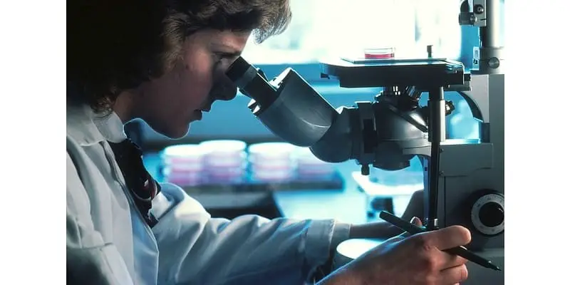
Biological Inverted Microscope
Biological microscopes are generally designed to be smaller than their industrial counterparts, just large enough to accommodate biological samples in petri-dishes or flasks. Slide preparations required in upright microscopes alter the pressure, oxygen content and temperature in the cell sample, thus significantly altering their activity and structure.
Using an inverted microscope, cell cultures can be magnified in their natural state in the medium. While petri-dishes can be accommodated in a traditional microscope, you risk the objective dipping into the culture medium and contaminating the sample. Furthermore, infectious organisms like fungal specimens can be magnified without opening the petri dish.
Biological inverted microscopes generally range between 40X to 400X magnification. These are also combined with high-resolution cameras to capture images of the magnified sample. Fluorescent inverted microscopes are the most common devices used in biology labs to examine distinct cellular structures.
Although traditionally cells needed to be dyed with fluorophores to be visualised, nowadays, cells can be genetically modified to secrete their own fluorescent dyes. Inverted microscopes have also found use in hospitals for live-cell observation and in-vitro fertilization.
If you think you could benefit from the advantages that an inverted microscope can provide Amazon actually has some solid but pricey biological inverted microscopes.
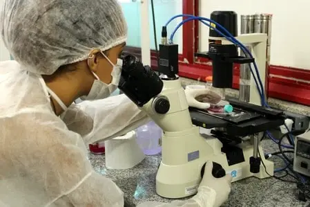
Metallurgical Inverted Microscopes
Metallurgical inverted microscopes can be used to examine specimens that are too heavy or large to be accommodated into an upright microscope. Since these devices do not require sample preparation, a large number of samples can be quickly examined in an industry, allowing for high throughput production.
Small tools can also be added to the device, to allow inverted microscopes to be used in micromanipulation applications. Currently, these devices have found use in manufacturing processes, quality check (fatigue, corrosion, cracks, ruptures), grain sizing, electroplating, telecom and electronics manufacture and watch production.
What are the Disadvantages of the Inverted Microscope
- Cost: Inverted microscopes that can provide good resolution and magnification are generally costlier than their upright counterparts. This is due to the complexity of the design.
- Container Aberration: The type of container used to house the sample can affect the refraction of light and thus the quality of the image.
- Large Working Distance: The large working distance is an advantage in certain cases, but it also limits the quality of illumination devices and condensers that can be used. The maximum magnification allowed in these devices is also much more limited than upright microscopes.
Takeaways
Inverted microscopes have some distinct advantages over their upright counterparts. The ability to view specimens without staining or introducing contaminates has allowed scientists to view some of the most incredible microbiological functions and processes at high magnifications.
If you are working with live specimens or specimens that you need more space with or a greater working distance you should definitely consider the advantages that you might get over a traditional upright microscope.
References
- Development of Low-Cost Inverted Microscope to Detect Early Growth of Mycobacterium tuberculosis in MODS Culture
- An Inexpensive Inverted Microscope System
- Flexible microassembly methods for micro/nanofluidic chips with an inverted microscope
- Under the Microscope: A Brief History of Microscopy
- History of the Optical Microscope in Cell Biology and Medicine