
A compound microscope is a microscope that utilizes a system of compounding lenses that enables the microscope to produce highly magnified images. Some of the lenses involved in this compound lens structure are the condenser lens, objective lens (which are themselves made up of several lenses), and the eyepiece lens. Compound microscopes can produce images magnified anywhere from 40x – 2,500x.
There are many types of microscopes, in fact you can check out Types of Microscopes: A Complete Breakdown to see them all, but the compound microscope is among the most recognizable because it is arguably the most common microscope used.
What are Compound Microscopes Used For?
- Compound microscopes are used in the medical industry to diagnose disease and to conduct many different types of experiments and research in various fields of study.
- Pathologists use compound microscopes to examine tissue and detect the existence of cancer. Compound Microscopes are used by forensic scientists to analyze and collect evidence for law enforcement.
- They are used by botanists to study plant development and microorganisms that interact with various plant species.
- Jewelers and gemologists used compound metallurgical microscopes to examine the structure and complexities of various gems and jewels to aid in appraisals.
- Geoscientists and Environmental Scientists use compound microscopes to examine water and soil samples for pollutants and foreign bodies.
- Compound microscopes are also used to examine nanocircuits and other extremely small electronics.
What Are Some Types of Compound Microscopes?
Here is a list of some of the most common compound microscopes.
- Biological Microscope
- Phase Contrast Microscope
- Polarizing Microscope
- Metallurgical Microscope
Now let’s take a closer looks at each one of these types of compound microscopes.
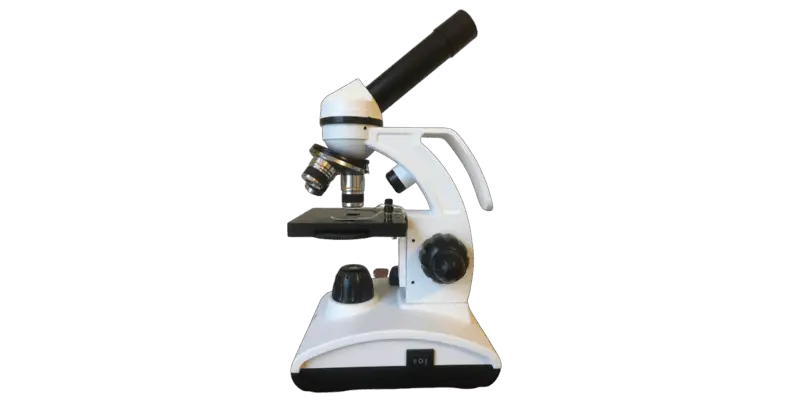
Biological Microscope
A brightfield microscope is what people commonly call a “Cell Microscope” or a “Biological Microscope”. This type of compound microscope is really more of a method of microscopy where light from a light source is channeled and focused up through a condenser lens as a cone of light and the refracted light through the specimen is what forms the image that makes its way through the magnifying lenses of the objective, through eyepiece lens and into the eye of the observer.
The sweet spot for this type of compound microscope are specimens that are not highly transparent but where light is able to refract and bend and bounce through and around the specimen. This is the microscope I have and it is a really great microscope to get into microscopy as a hobby. It is not prohibitively expensive and it has everything you need including a nice camera for video and photos.
Phase Contrast Microscope
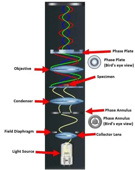
A phase contrast microscope is also not really a microscope but more of a method of microscopy that uses a compound microscope. With a phase contrast annulus condenser attachment and phase contrast objectives and some specific configuration, you can turn a brightfield microscope into a phase contrast microscope. This kit contains everything you need to turn your standard brightfield microscope into a phase contrast microscope.
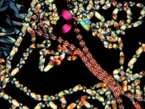
Phase contrast is useful when you have a highly transparent specimen and you do not want to stain the specimen because it will either kill it or interfere with it in some way. Because the light passes through the specimen with very little refraction it makes certain details very hard to visualize. Phase contrast microscopy works by first by sending light from the light source through the annulus ring which has three curved slits (as shown in the image). For more on phase contrast microscopy check out this post.
This creates ringed light that passes up through the condenser which then either passes straight through the specimen or is diffracted by biological structures or parts of the specimen. The light that passes straight through the specimen goes through the objective and slowed down by a phase plate that is positioned at the back of the objective lens. Without getting too technical this relative change or slowing of the space, diffracted, and particle waves of light are what produce the contrast that the observer sees. This contrast is what enables the observer to see details that would be all but invisible under a brightfield configuration.
Polarizing Microscope
A polarizing microscope is built with a polarizer and an analyzer that is used to pick up light differences after the light is polarized, refracted through the specimen which then makes the light waves become out of phase.
The light waves are then recombined through the analyzer. This enables the microscope to detect information about absorption of color and optical path boundaries.
Metallurgical Microscope
Metallurgical microscopes work on based on reflected light instead of refracted light. Light information is gathered by the objective from the reflected light back into the objective rather than the light going up and refracting through the specimen like in a biological microscope.
What this means is that typically the types of materials that are observed are these microscopes are highly dense materials where light does not pass through the specimen. Stereo microscopes also use this refracted light principle, but they often cannot achieve the magnification levels that a metallurgical microscope can.
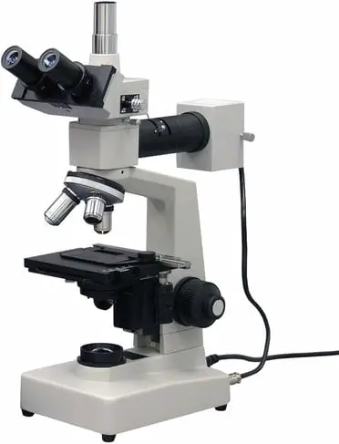
Metallurgical microscopes often come with a darkfield option that can make detecting anomalies and fractures in certain materials easily detectable.
What are the Limitations to Compound Microscopes?
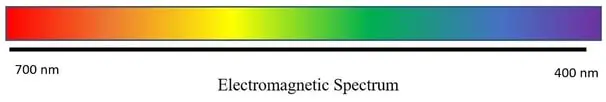
The two main limitations to compound microscopes are light and working distance. Although compound microscopes can achieve very high magnifications (up to 2,500x) some specimens are so small that they still are not visible even under these extremely high magnifications. This is because the ability for light to refract hinges on the wavelength of light being smaller than the specimen. When you start to try to visualize specimens that are smaller than the wavelength of light, compound light microscopes will fail to produce an image.
For example, a compound biological microscope would be able to image a bacterium successfully but not a virus. A bacterium is around 1 micrometer and a virus is around 0.02 micrometers. The smallest wavelength of visible light is 0.4 micrometers. For more on electron microscopes see Electron Microscopes Explained: From Physics to Images.
Specimen Space and Working Distance
Because the compound microscope achieves very high magnifications it often requires very small working distance between the specimen and the objective lens.
This limits the type of specimen that can be observed. For larger specimens at lower magnifications a digital microscope or hand held microscope may be more useful. For slightly larger specimens that will not fit on an upright compound microscope, an inverted microscope may be more suitable.
Who Invented the Compound Microscope?
If you take the definition of a compound microscope as using a device with more than one lens to magnify an object image, than you may credit the great Galileo Galilei (1564 – 1642) as the inventor of the compound microscope. After rearranging the lenses in his telescope, he was able to magnify objects. The history of the compound microscope is a story of scientific triumph, but it is not without some debate. Depending on how you define the compound microscope, different historians may credit different inventors.

If you define the invention of the compound microscope as more closely resembling the designs of compound microscopes of today, you may credit Zaccharias Janssen (1585 –1632~) as the inventor of the compound microscope. Both men lived around the same time and came up with their designs completely independently of each other. For the complete history of microscopes see The First Microscope to Modern Microscopes.
How Much Does a Compound Microscope Cost?
Compound microscopes cost on average around $900-$1,200. The cost of a compound microscope can vary depending on the quality, features and brand. For a more detailed breakdown of microscope prices check out this article.
It can seem like a pretty steep investment but depending on your level of interest in microscopes and microscopy there is a compound microscope out there for you and for what you are trying to explore. Keep in mind like with most things, you get what you pay for. This is especially true in scientific instruments like microscopes, where quality can be the difference from a stunning image and a blurry mess.
How Much Does a Compound Microscope Weigh?
Compound microscopes can range in weight from 3 pounds for a smaller compound microscope and up to 20 pounds for a lab grade microscope with a mounted camera. The weight of a microscope is important in a lab setting where minute disturbances to the position of the microscope can cause a specimen to be moved from the field of view or focus to be diminished. Smaller lighter compound microscopes are better for field observations where the microscope will be carried to a remote location away from a traditional lab.
Takeaways
The compound microscope is an incredible scientific instrument used to develop medicines, for quality assurance in high technology fields, and to discover disease and pathogens. These highly complex and precise instruments can open up the microscopic world to people new to microscopy and provide new insights and discoveries to seasoned professionals.
