

Spirilla refers to a group of bacteria found in the family Spirillaceae. They are classified as spiral-shaped, aerobic, gram-negative bacteria with flagella (thread-like appendages that allow movement). Spirilla are found in aquatic environments rich in organic matter. Spirilla are long and thin in shape, making them efficient for effective nutrient uptake due to their large surface-to-volume ratio.

.jpg)
How Are Spirilla Bacteria Classified?
Spirillum was first documented in the late 1600s. The genus Spirillum was formed by Gottfried Ehrenberg, a German scientist in 1832. At that time, the genus contained many species of aerobic and microaerobic bacteria as well as chemoheterotrophic bacteria.
In 1973, the characteristics of 39 strains of Spirillum were assessed and a paper by Hylemon et al. was released which resulted in the reorganization of the genus Spirillum into 3 separate genera. They were separated by their percentage molecular DNA base composition and by their physiological characteristics.
The 3 resulting genera were Aquaspirillum, Oceanospirillum, and Spirillum. Each of these genera will be discussed in detail below. Even though the genus Spirillum has been split into 3 distinct genera, the term Spirilla (plural) or Spirillum (singular) is still used to describe any helical or spiral-shaped bacteria.
Genus Spirillum
Firstly, we will look at the Genus Spirillum. Bacteria in this genus are found in the phylum proteobacteria, the class Betaprotobacteria, and the order Nitrosomonadales. Their family is Spirillaceae. There are 3 species currently in the genus Spirillum. These are S. volutans, S. winogradskyi, and S. Kriegii. However, S. minus is sometimes placed in this genus. Originally named back in 1924, S. minus is not on the Approved List of Bacterial Names. This is because it does not have an identified reference strain. Therefore, technically does not belong in this genus.
The genus Spirillum contains freshwater bacteria that are large and only require a small amount of oxygen (microaerophilic) to survive. This is usually a percentage of around 1-9%. The size of Spirillum volutans is approximately 5 – 8 micrometers wide and 60 micrometers long. Spirilla have a spiral or corkscrew-like appearance. Their structure is rigid due to their rigid cell wall.
This differs from spirochetes from the Phylum Spirochaetes which have a flexible cell wall allowing movement. Spirilla contain flagella which are filamentous protein appendages that aid in movement. This allows them to be mobile as bacteria in this genus are usually found in stagnant water. Spirilla contain volutin granules which are composed of poly-β-hydroxybutyrate. Initially named volutin granules in 1895 because of their discovery in Spirillum volutans, they are also present in many other microorganisms, not just Spirilla.
Volutin granules are inclusion bodies of poly-β-hydroxybutyrate. They serve as energy stores in the cytoplasm of the bacterial cells. These granules can be stained by toluidine blue or methylene blue and then viewed under the microscope. The granules are visualized as blue/black, distinct against the color of the bacterial cells.
Structure Of Spirillum Volutans
Spirillum volutans have flagella which are lophotrichous in nature i.e. it has multiple flagella arising from one point. In this case, from both poles of the bacteria. The flagella rotate to allow motion. The bacterial cells can change their direction of movement by switching the motor rotation to the other direction. Originally it was thought that there was a single or only a few flagella at each pole subsequently, it was determined that they stick together during staining to look like one or a few flagella.
For example, S. volutans has around 50 flagella at each pole. A loss of rotation and movement can be seen when there is a change in the environmental conditions, for example, toxicants such as heavy metal ions. The motility of Spirilla allows them to escape predators and internalization by other organisms.
Flagella also have the function of acting as mechanoreceptors and can respond to connections with surfaces. The cells do not adhere via the flagella but by the tip of one of the poles of the cell.
The growth of S. volutans on solid media is difficult. Its growth is best achieved by culturing in a peptone-succinate-salts broth in micro-aerobic conditions. However, it is possible to grow aerobically when the peptone is replaced by a vitamin-free acid-hydrolyzed casein broth. Also, it can be grown aerobically if potassium metabisulphite, norepinephrine, and superoxide dismutase are added to the peptone-succinate-salts broth.
Genus Oceanospirillum
Oceanospirillum is another genus consisting of spiral-shaped bacteria originally place in the genus Spirilla. This group of bacteria is found in marine environments in the Phylum Proteobacteria. They are from the Class Gammaproteobacteria, the order Oceanospirillales, and their family is Oceanospirillaceae. They are gram-negative and like Spirillaceae, are helical in shape with a few exceptions. The optimum temperature for their survival is 25 – 32°C and seawater is necessary for growth to occur.
Bacteria in this genus have a DNA molecular GC percentage (Guanosine-Cytosine) of around 42-48%. This is higher than the DNA molecular CG percentage found in the genus Spirillum, thus separating them into separate genera.
Their helical shapes have a clockwise direction, and their flagella can be located at both (amphitrichous) or just one (monotrichous) of the bacterial poles. Oceanospirillum is small in diameter (0.4 – 1.4 micrometers) and their length is between 1.2 and 75 micrometers. They are aerobic chemoheterotrophs and oxygen is their terminal electron acceptor.
Some species in the genus Oceanospirillum are rod-shaped, rather than spiral shaped. There are still proposals that Oceanospirillum should be further classified, as currently, it is taxonomically heterogeneous.
It is also worth noting that changes in the shape of the bacteria can occur when some species are cultured for prolonged periods. For example, during the prolonged culture of O. japonicum, the cells become less helical and have only an S-shape or a slight curve to them.Some examples of Oceanospirillum are O. japonicum, O. beirjerinckii, and O. minutulum. The full list can be seen in the table below. Oceanospirillum can be isolated from coastal seawater, as well as rotting seaweed and the insides of rotten marine mussels.
| Oceanospirillum |
|---|
| O. beijerinckii |
| O. commune |
| O. hiroshimense |
| O. jannaschii |
| O. japonicum |
| O. kriegii |
| O. linum |
| O. maris |
| O. minutulum |
| O. multiglobuliferum |
| O. nioense |
| O. pelagicum |
| O. pusillum |
| O. sancuarii |
| O. vagum |
Oceanospirillum has a polar membrane usually located on the areas that contain the flagella. It is connected to the inside of the plasma membrane via bridges. This genus also contains volutin granules of poly-β-hydroxybutyrate for energy storage. If cultured for prolonged periods, Oceanospirillum forms coccoid bodies.
These are also known as microcysts. When the bacteria join to form coccoid bodies, their cell walls become very thin. The process occurs when one cell meets another cell, and they entwine together. Here, they fuse and over time become smaller and rounder, resulting in a coccoid body. In nutrient seawater agar, colonies generally form within a few days.
The colonies are white, pale yellow, or yellow-green depending on the species and have a size of around 1.5mm in diameter. If the coccoid body is placed in fresh media, the cells can grow back to a helical shape, resulting in their ‘germination’.
Genus Aquaspirillum
Aquaspirillum is also a spiral-shaped bacteria which are frequently found in stagnant water, just like Spirillum. Aquaspirillum is also gram-negative, rigid, helical cells with the exceptions of Aquaspirillum delicatum which is a curved rod shape, and Aquaspirillum fasculus which is a typical rod-shaped bacterium. The bacteria in this genus also contain flagella or tufts as is found in the genera Oceanospirillum and Spirillum. They vary on their presence and quantity of flagellum. One species may have only a single flagellum at one pole (monotrichous), whereas others may have a single flagellum at each pole (amphitrichous).
Taxonomically, they are found in the phylum proteobacteria, the class betaproteobacteria, and order Neisseriales due to their differences from Spirillum and Oceanospirillum species. For example, Aquaspirillum describes freshwater bacteria that do not tolerate high levels of salt (3% or above) and have an even higher DNA molecular base GC percentage content than Oceanospirillum.
Aquaspirillum is usually aerobic although some have been found to survive in micro-aerobic conditions. They respire using oxygen as the terminal electron acceptor although nitrate may be used anaerobically in some species. The optimal growth temperature for this genus is around 30°C. A few species can utilize carbohydrates, but they are not usually metabolized. Instead, they tend to use amino acids or the salts from organic acids as their sources of carbon.
Species in this genus have a diameter of between 0.2 and 1.4 micrometer. They are described as chemoorganotrophic which means that they obtain energy from chemical bonds in oxygen or organic compounds. This results in the oxidation of organic compounds such as fats, sugars, and proteins. However, one species, Aquaspirillum autotrophicum can oxidize hydrogen and is therefore facultatively autotrophic.
As there is quite a broad range of differences between species in this genus, a study in 2006 suggested that only 3 species are truly Aquaspirilla. These are Aquaspirillum serpens, A. bengal, and A. fasciculus. However, to date, the genus has remained unchanged due to difficulties in separating the highly heterogenous species which contains over 10 species of bacteria shown in the table below. An example of a bacterial species in the genus Aquaspirillum (A. Bengal) will be described in more detail below.
| Genus Aquaspirillum |
|---|
| A. anulus |
| A. aquaticum |
| articum |
| autotrophicum |
| bengal |
| delicatum |
| dispar |
| fasciculus |
| giesbergeri |
| gracile |
| itersonii |
| magnetotacticum |
| metamorphum |
| peregrinum |
| polymorphum |
| psychrophilum |
| putridiconchylium |
| serpens |
| sinuosum |
| soli |
Aquaspirillum Bengal
Aquaspirillum bengal was initially isolated from West Bengal in a freshwater pond. It has a DNA base composition of 51% GC. It has a diameter of between 0.9 micrometers and 1.2 micrometers and a cell length of between 5.2 – 22 micrometers. A. bengal has a similar diameter to A. serpens and A. putridiconchylium. It also has a very high temperature of growth, showing optimal survival and growth rates at 41°C. It has an optimum pH of between 6 and 8.4.
In microbiological culture, the colonies of A. bengal are light brown. The bacteria have a swimming speed of between 40 to 52 micrometers per second. A. bengal thrives in microaerophilic conditions but cannot tolerate salt of greater than 1%.
How To Observe Spirilla Under The Microscope?
Spirilla can be seen under a standard light microscope using phase contrast microscopy techniques or closing the iris diaphragm to a position where it is only slightly open.
Diseases Caused By Spirilla Bacteria
Spirillum minus, although technically not in the genus Spirillum, causes rat-bite fever. This disease is most commonly found in Asia. Rat-bite fever can also be caused by different bacteria, Streptobacillus moniliformis. Infection occurs after being bitten by a rat. Around 1 – 4 weeks after the bite, the individual will develop a fever, headache, and pain at the site of the bite. Ulcers may occur at the site of injury, which is painful and prevent healing.
A rash may appear which will resolve once the patient has recovered. If the disease is left untreated, symptoms can last up to 8 weeks in recurring cycles. Worst cases result in pneumonia, meningitis, myocarditis, or endocarditis. Treatment of patients suffering from rat-bite fever is with the antibiotic penicillin. Penicillin gives an excellent disease outcome, but Streptomycin and tetracycline can also be used if Penicillin cannot be used.
What Are Spirillum-Like Bacteria?
As mentioned above, S. minus has never been properly identified. It is associated with rat-bite fever, a disease transmitted by rats or through ingestion. As other species of the family Spirillaceae are usually obligately microaerophilic and not found in mammals, this further supports the notion that this species of bacteria were assigned to the wrong genus.
Although S .minus has not yet been grown on artificial media, it can be seen on darkfield microscopy or in Wright or Giemsa stained blood smears.
What Are Spirochetes?

Spirochetes are also gram-negative bacteria like Spirilla. They are also mobile and found in freshwater environments. Although they have many similarities with Spirilla, they are not classed as Spirilla bacteria. They have a flexible cell wall, in comparison to spirilla, which have a rigid cell wall. Spirochetes are long, up to 250 micrometers with a diameter of between 0.1 and 3 micrometers. Spirochetes have a double membrane that consists of a bacterial cell wall enclosed by an outer membrane.
The periplasm, the narrow space between the inner and outer membrane contains periplasmic flagella (endoflagella). The flagellum are attached to a motor that is connected to the inner membrane. They are also connected to the cell wall at each polar end of the bacterium. In this way, they wrap inside and along the cell body. They can have between 1 and 100 periplasmic flagella which aid in movement but also have a skeletal function.

Spirochetes are found as free-living bacteria occurring naturally in water, but they can also be symbionts of insects or parasites in animals.
Spirochetes are classified based on their pathogenicity and morphology. They are difficult to culture and because they are so slender, they are also difficult to view under a microscope. Spirochetes are placed in the order Spirochetales which consists of two families, Spirochaetaceae and Leptospiraceae.
There are four genera in Spirochetaceae which include Spirocheta, Cristispira, Treponema, and Borrelia. Spirocheta and Cristispira are found free-living or commensally in organisms (live with the animal or insect without causing harm). Pathogenic species can be found in the genera Treponema, Leptospira, and Borrelia.
Spirochetes are found in a wide range of environments, from soil and water to decaying matter and in plants and animals. They can be viewed by dark microscopy or phase microscopy or by staining with Giemsa or silver impregnation. Borrelia can be stained with aniline dyes.
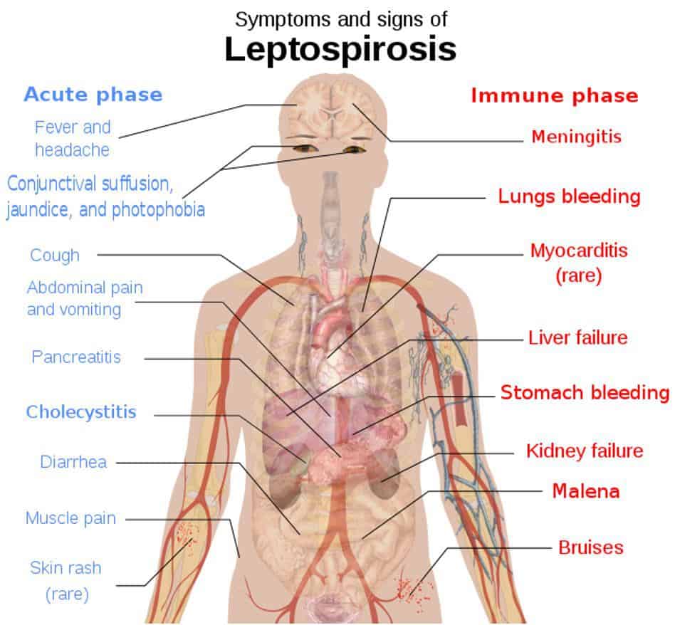
Some examples of spirochetes include Borrelia burgdorferi, which causes Lyme disease, and Treponema pallidum which causes Syphilis. Leptospira interrogans is another infectious spirochete that causes leptospirosis.
Borrelia Burgdorferi
Borrelia burgdorferi is part of the spirochete class of bacteria that causes Lyme disease. This can affect both animals and humans, it is spread by ticks in woodland areas. Ticks carry B. burgdorferi, and in fact, have co-evolved with them over millions of years. The tick must be attached to the animal for at least 24 hours for the bacteria to be transmitted to the host. Ticks become infected when they bite an infected animal or human. They can then pass it on to their next victim through the bloodstream.
B. burgdorferi can affect any part of the body, such as the brain, muscles, joints, or even the heart. It is difficult to diagnose due to the wide range of symptoms it gives. Therefore it is common to be misdiagnosed with fibromyalgia, MS, fatigue, or depression. Lyme disease is found everywhere except Antarctica. Thankfully, it can be successfully treated with antibiotics.
Leptospira Interrogans
The bacteria Leptospira interrogans is the Spirochete responsible for causing Leptospirosis. The bacteria is found in many animals and carried in their kidneys. It is more commonly found in tropical areas. Left untreated, leptospirosis can cause a severe illness known as Weil’s disease which has severe symptoms of renal failure, pulmonary hemorrhage, and jaundice. It can even cause death.
The disease can be treated with penicillin in severe cases, or in mild cases, it can be treated with doxycycline. There is no current vaccine available commercially for humans although doxycycline can be used as a prophylactic (preventative) treatment in areas where there are outbreaks. Thankfully, most people, only suffer a mild illness from Leptospira interrogans.
Leptospirosis spreads from animals with chronic infections of the renal system. This is usually by mammals, but reptiles and amphibians are also able to carry the bacteria. The infected urine is then spread into the environment. Humans can become infected through contaminated soil or directly through an infected animal. The bacteria enter the body through cuts in the skin or via the mucous membranes. Here, they can enter the bloodstream.
The bacteria can move using their flagella and attach to endothelial cells of the blood vessels and the extracellular matrix. They spread throughout the body through the bloodstream and cause damage to the liver by killing liver cells. This in turn can lead to jaundice. The toxins produced by Leptospira interrogans are responsible for causing kidney damage.
Spirilla vs Spirochetes
It would be easy to confuse Spirochetes, found in the order Spirochetales with Spirilla due to their spiral shapes. However, many Spirochetes cause harmful diseases in humans, unlike Spirilla. They have endoflagella (or periplasmic flagella) which vary in number depending on the species, this differs from Spirilla which has external flagella.
As mentioned above, the endoflagella are wrapped around the bacteria forming an axial filament. The Spirochetes move using the rotation of the endoflagella, resulting in a twisting, forward motion. Table 3 shows the differences between Spirilla and Spirochetes.
| Spirilla | Spirochetes |
|---|---|
| Cell wall: Rigid | Cell wall: Flexible |
| External flagella | Internal flagella |
| Size > 1 µm | < 1 µm |
| Spirilla = genera | Spirochetes = phylum |
| Gram negative | Difficult to gram stain |
| Aerobic | Obligate or facultative anaerobes |
| Disease example: rat bite fever | Disease example: Lyme disease or Syphilis |
References
- Aryal, S. (2018). Different size, shape, and arrangement of bacterial cells. Microbiology info.com. https://microbiologyinfo.com/different-size-shape-and-arrangement-of-bacterial-cells/
- Aquaspirillum Bengal. Microbe Wiki. Aquaspirillum bengal – microbewiki (kenyon.edu)
- Cole, J.R. (1990) Spirochetes. Diagnostic Procedure in Veterinary Bacteriology and Mycology (Fifth Edition), 41-60. https://doi.org/10.1016/B978-0-12-161775-2.50009-8
- External Structures of Prokaryotic Cells. (2016). Science Prof Online. https://www.scienceprofonline.com/cell-biology/external-structures-prokaryotic-cells.html
- González J.M., Whitman W.B. (2006) Oceanospirillum and Related Genera. In: Dworkin M., Falkow S., Rosenberg E., Schleifer KH., Stackebrandt E. (eds) The Prokaryotes. Springer, New York, NY. https://doi.org/10.1007/0-387-30746-X_33
- Hylemon, P.B., Wells, J.S., Krieg, N.R. Jannasch, H.W. (1973). The Genus Spirillum: A Taxonomic Study. International Journal of Systematic Bacteriology. 23(4) 340-380. https://www.microbiologyresearch.org/docserver/fulltext/ijsem/23/4/ijs-23-4-340.pdf?expires=1606397807&id=id&accname=guest&checksum=C0CB13F9B7639B83E1ADF96FDD9A137D
- Krieg, N.R. (1976) Biology of the chemoheterotrophic Spirilla. Bacteriological Reviews. 40(1) 55-115. https://mmbr.asm.org/content/mmbr/40/1/55.full.pdf
- Krieg N.R. (2006) The Genus Spirillum. In: Dworkin M., Falkow S., Rosenberg E., Schleifer KH., Stackebrandt E. (eds) The Prokaryotes. Springer, New York, NY. https://doi.org/10.1007/0-387-30745-1_29
- Nanninga, N. (2014). Cell Structure, Organisation, Bacteria, and Archaea. Reference Module in Biomedical Sciences. https://doi.org/10.1016/B978-0-12-801238-3.02309-6
- Nazir, R., Rehman. S, Nisa, M., Baba, U. (2019). Exploring bacterial diversity: from cell to sequence, Freshwater Microbiology, Academic Press. (7) 263-306. https://doi.org/10.1016/B978-0-12-817495-1.00007-4
- Pot B., Gillis M., De LEY J. (2006) The Genus Aquaspirillum. In: Dworkin M., Falkow S., Rosenberg E., Schleifer KH., Stackebrandt E. (eds) The Prokaryotes. Springer, New York, NY. https://doi.org/10.1007/0-387-30745-1_30
- Shively, J.R et al. (2009). Intracellular Structure of Prokaryotes: Inclusions, Compartment, and Assemblages. Encyclopedia of Microbiology. Third edition pages 404-424. https://doi.org/10.1016/B978-012373944-5.00048-1
- Vinetz, J.M., Watt, G. (2020). Leptospirosis. Hunter’s Tropical Medicine and Emerging Infectious Diseases. (10th Edition) 636-640. https://doi.org/10.1016/B978-0-323-55512-8.00079-X
- Wolgemuth C. W. (2015). Flagellar motility of the pathogenic spirochetes. Seminars in cell & developmental biology, 46, 104–112. https://doi.org/10.1016/j.semcdb.2015.10.015