

It’s easy for us to forget about the complex and diverse ecosystem that lives right under our noses. Without the help of a microscope, the human eye is unable to observe the many microorganisms that live amongst us. Vorticella are just one of many microorganisms that live in this otherworldly environment.
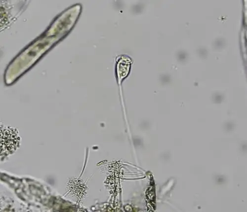
Vorticella are microscopic bell-shaped freshwater aquatic organisms. These heterotrophic and predatory organisms are mostly sessile. Vorticella are identified by their bell-shaped bodies and long stalks that attach to a substrate. The body ranges from 30 to 40 micrometers while the stalk can grow up to 100 micrometers in length.
Most Common Types of Vorticella
There are approximately 177 species of vorticella. Of those 177, the most common species and their traits are listed below:
- Vorticella campanula: This species is the most common amongst the Vorticella and most heavily studied. The campanula species are distributed worldwide and are not colonial nor solitary but social. Social meaning that several can be found together at a time. This species is found mostly in stagnant water with decaying organic matter that they can feed on.
- Vorticella citrina: All other species of vorticella, including the citrina have been lightly studied. This species in particular is known for its yellow to light green color.
- Vorticella marina: This species has adapted to live in shallow water. They can only be found at a depth of up to one meter. This means that they are mainly found in bays and near the shoreline.
- Vorticella parapulchella: This species is distinguished by its tiny size and widely spaced pellicular ridges. They are also known for having a highly unusual infundibular polykinety. In particular, they only have two rows instead of three.
- Vorticella fusca: Not much is known about this species, although it has a unique oral infraciliature and pellicular morphology.
Since many of the vorticella species are not well studied, their characteristics are not widely available to the general public, especially for students. There are many published studies on these species, but may be difficult to digest for a student. Much of the information from the studies have not yet been summarized and published on blogs or websites; therefore, most of the information is hidden from the general public.
What do Vorticella Eat?
As mostly sessile organisms, vorticella have adapted to become suspension feeders. The vorticella’s buccal cavity, or its mouth, is surrounded by cilia. Cilia are responsible for catching suspended food sources and directing them towards its mouth and into its digestive cavity.
Oral cilia beat and vibrate and to create an inward flow of water which ultimately pulls food closer. The current created is comparable to a mini vortex. It’s hypothesized that the contractile in the stalk also helps the vorticella create a current, or otherwise expand and contract to access different food sources.
Vorticella’s diet mainly consists of bacteria and small flagellates. In reality, as suspension feeders, they are not picky and will eat just about anything that flows through their peristome. If the food supply is scarce, vorticella have the ability to detach and relocate in search of a more abundant area. Telotroch, on the other hand, does not feed. In this early stage of life, telotroch utilize their somatic cilia as an energy source, which they use for growing a stalk and undergoing metamorphosis.
Structure and Anatomy of Vorticella
- Double row of cilia: Cilia are the fine, hair-like structure found at the peristome, or mouth opening, of the vorticella. This structure has the ability to pulse and vibrate, effectively creating a current that pulls food towards the peristome. The cilia are also able to grab onto food and navigate it towards the peristome.
- Cilia membrane: The cilia membrane is not to be mistaken with a plasma membrane, although it is continuous with the plasma membrane. The cilia membrane differs in that it is made up of unique lipids and proteins.
- Peripheral shelf: The peripheral shelf is the layer to which the cilia are attached. It’s a rigid and donut-shaped structure that is layered around the peristome.
- Buccal cavity: The buccal cavity, or otherwise known as the mouth, is a funnel-shaped structure that is the beginning of the digestive process.
- Contractile vacuole: The contractile vacuole is located next to the macronucleus and the vestibule. It is a complex organelle that is unique to free-living organisms. It regulates and keeps a balance of intercellular water. It can either accumulate or expel water to keep this balance. This structure helps the vorticella create an inward current and also helps it expel unwanted foods that get trapped in the buccal cavity.
- Macronucleus: The macronucleus is a uniquely large nucleus found in ciliates. It is responsible for all metabolic activities. During sexual reproduction, the macronucleus breaks down and is responsible for organizing the genetic material.
- Spasmoneme: Otherwise known as a myoneme, the spasmoneme is the contractile structure responsible for the stalk’s ability to contract and retract. It is made up of proteins that quickly shorten when exposed to calcium. This structure is particularly amazing because it can contract within milliseconds, much quicker than the blink of an eye. It can then extend itself within another two seconds. It’s hypothesized that this structure allows the vorticella to retreat when threatened and may also help create an inward current/vortex to attract suspended food.
- Stalk sheath: The stalk sheath is the protective covering of the stalk. It is firm enough to keep it rigid while expended but flexible enough to coil and contract.
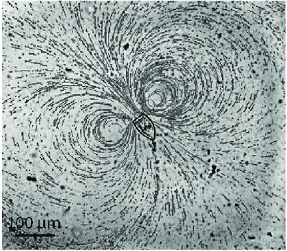
This photo illustrates the inward flow of water created by the cilia and contractile vacuole. The black lines represent the flow path of water that is created. Note that it resembles a vortex. It’s also interesting to note that there are two vortexes formed, each flowing in opposite directions. This photo represents the power of the vibrations that are created by the cilia. Although small in size, they work in sync to create a vortex that is strong enough to lure in potential food sources.
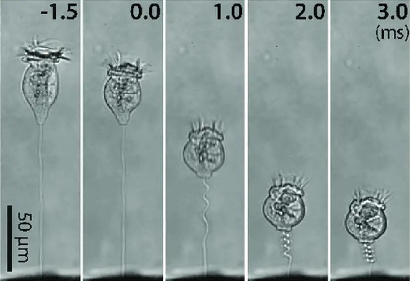
The leftmost photo represents the vorticella in an overextended ‘reaching’ phase. The next photo over shows the vorticella in a completely relaxed phase. The next three photos show the vorticella as it contracts. Note that the vorticella is able to stay suspended in any of the five phases at any given time. Also note that in the rightmost photo, the bulb is more circular than bulb-shaped. In this phase, the contractile vacuole has expelled its contents to be as small and compact as possible. It’s theorized that the vorticella contracts completely when it is threatened. It attempts to reduce its size to retreat from any potential harm.
Where do Vorticella Live?
Vorticella can most commonly be found in lakes, rivers, streams, and any other sources of fresh water. Some species can tolerate brackish or salty waters. Since they are mostly sessile, you will find them attached to some sort of substrate for their entire lives.
They are most commonly found attached to other aquatic vegetation. Attaching themselves to vegetation usually means that there is a plethora of food around them such as algae to sustain themselves. Vorticella can also be found attached to rocks, soil, algae, or even other animals.
It’s not uncommon that vorticella are found attached to crustaceans such as crabs or on the shells of mollusks. Attaching themselves to another living creature increases its chances of having easy access to other food sources. Vorticella can be found as either solitary individuals or within colonies. They are most commonly found amongst a colony. The colonies are not true colonies in the sense that each individual has its own stalk and it can leave the colony at any time. In other words, the vorticella is not dependent upon its colony for survival.
How do Vorticella Reproduce?
The vorticella is unique in that it can produce both asexually as well as sexually. Below we will explore the two different modes of reproduction and what makes them so adaptable.
Vorticella Asexual Reproduction
The more common of the two, asexual reproduction means that gametes are not necessary to reproduce. The vorticella does not need to rely on gametes or a partner to reproduce. Instead, it is capable of reproducing utilizing binary fission, or division.
Binary fission occurs when an organism splits into two daughter cells by means of propagation from the parent vorticella. In preparation for binary fission, the vorticella shortens in size and elongates. The peristome, or the mouth of the bell, closes. The contractile squeezes and releases in a pulsing pattern during binary fission.
The division of the nucleus shortly follows. The macronucleus separates amitotically; whereas, the micronucleus separates mitotically. The division is unique in the vorticella in that the two new individuals are unequal in size; whereas, most other organisms that undergo binary fission end up equal in size.
It’s also important to note that vorticella undergo binary fission longitudinally. Think of it like this – the vorticella split like a hot dog bun when it undergoes binary fission, whereas most other organisms split like a hamburger bun.
The larger of the two new organisms retain the stalk. The smaller organism will have to develop new cilia and grow a new stalk of its own so that it can attach to a substrate. The smaller organism is typically more cylindrical in shape and is referred to as the telotroch. As time goes on, the cylindrical shape will grow into the recognizable bell-shape. The telotroch swims away using its aporal cilia and finds a place to anchor.
The end product results in two genetically identical and fully independent vorticella. Amazingly, the entire process of division only takes 20 to 30 minutes. Vorticella are amongst the quickest when it comes to generating cellular and molecular movement.
Vorticella Sexual Reproduction
Vorticella are also capable of producing by means of conjugation. Conjugation happens when genetic material is transferred to another organism by means of direct contact. One of the organisms is responsible for donating genetic material, whereas the other organism serves as the recipient. This process is also known and sexual reproduction.
In vorticella, conjugation takes place in two separate phases. The first phase consists of the formation of micro and macro-conjugants. The phase takes advantage of binary fission, similar to how vorticella asexually reproduce. The result of the binary fission is one large and one small cell.
The small cell is known as the micro-conjugant. Micro-conjugants are extremely similar to telotrochs in that they develop their own cilia, break away from the larger cell, and swim to a new surface where they anchor themselves. The main difference is that micro-conjugants are much smaller.
If the micro-conjugant does not come in contact with a macro-conjugant, then it will likely die within one day. Unlike a telotroch, micro-conjugants do not grow a stalk and do not undergo an encysting stage. Encysting is a process in which a telotroch encapsulates itself in a hard shell to protect itself from the environment.
The macro-conjugant, on the other hand, retains the stalk. In this phase, the macro-conjugant is fully capable of reproducing with other micro-conjugates.
The second stage of conjugation is called fission. Micro-conjugates use their cilia to swim around until they come in contact with a macro-conjugate. The micro-conjugates uses its cilia to attach itself to the stalk of the macro-conjugate. The macro-conjugate engulfs the micro-conjugate through fission and a zygote is formed. The nucleus of both conjugates undergoes a complex series of divisions. The end product of these divisions gives birth to seven daughter cells. The daughter cells undergo metamorphosis until they mature. Once matured, each cell becomes its own individual and self-sustaining vorticella.
When Were Vorticella First Discovered?
Vorticella were first identified by a Dutch businessman and scientist named Antonie van Leeuwenhoek. Leeuwenhoek first wrote about vorticella in a letter dated October 9, 1676. He wrote about a small creature with a pair of horns near its mouth which moved like a horse’s ears. It wasn’t until later that this feature was actually the vorticella’s oral cilia working to create a flow of water.
In 1755, a German painter named August Johann Rösel painted the first picture of the vorticella. Rösel was known at the time for his detailed paintings of microorganisms. Rösel had initially referred to the creature as Hydra convallaria. It wasn’t until 1767 that the organism was renamed Vorticella convallaria.
A scientist by the name of Otto Friedrich Müller claimed to have discovered 127 different species of vorticella in 1786. Many of his claims were later reversed as they were actually protozoans or rotifers. This is not surprising as vorticella physically resembles the phylum Suctoria. The name vorticella was given by Ehrenberg in 1838. Since then about 80 species of vorticella have been identified. Today, there are over 100 species that are recognized as vorticella by the biology community.
How Are Vorticella Classified?
The kingdom, phylum, class, order, and genus of the vorticella are described below:
- Kingdom: Chromalveolata – Proposed by Thomas Cavalier-Smith in 1981, the kingdom Chromalveolata is a supergroup that stemmed from a reorganization of Cavalier-Smith’s Chromista kingdom. It is now regarded as one of the six major groups of eukaryotes. The kingdom Chromalveolata consists of all of the organisms that descended from a single secondary endosymbiosis of a red alga and a bikont. This kingdom is contentious amongst the biology community as it was reorganized once more in 2006. The kingdom is considered a working group and is subject to change.
- Phylum: Ciliophora – Ciliophora come from a single Protista lineage. Otherwise known as ciliates, cilia are present in all ciliates at some point or another. Members of this phylum interestingly have two nuclei: a smaller genome-containing nucleus, and a larger one that helps with cellular regulation. Samples of this phylum are uncommon as it’s difficult to preserve a Protista when it lacks cell walls. Ciliophora as best observed straight out of their natural habitat, or even within their natural habitat if possible.
- Superphylum: Alveolata – The superphylum Alveolata consists of three main groups: the dinoflagellates, the apicomplexan parasites, and free-living ciliates. Half of the dinoflagellates are algae, most abundantly a large microalga. These algae are largely responsible for algal blooms. Apicomplexan parasites consist of just about all parasites in existence. Some of these parasites can cause diseases such as malaria and cryptosporidiosis in humans as well as many livestock diseases. Parasites are known for their invading a host and feeding off what the host has to offer. Vorticella are free-living ciliates which are commonly identified by their cilia. Cilia are virtually identical to eukaryotic flagella but are shorter in length and more abundant. About 45,000 free-living ciliates have been identified. Ciliates range from just 10 micrometers to 4 millimeters.
- Class: Oligohymenophorea – The class Oligohymenophorea consists of an abundant class of ciliates. Oligohymenophorea can be further identified by its ventral groove that makes up the mouth, which is separate from the body. A paroral membrane sits next to its mouth and typically has the membranelles to its left. The result of this is three brush-like structures that are used to undulate and create a flow of water with food and other nutrients towards the mouth. The food is then filtered through the cilia and ingested. This is how most ciliates in the Oligohymenophorea class get their nutrients. The class Oligohymenophorea was first identified in 1974. It is just one of three classes of ciliates. The composition of this class has remained consistent over the years. Only the apostomes have been added since.
- Subclass: Peritrichia – Otherwise known as the peritrich, this is a large subclass of ciliates. The subclass was initially proposed in 1859 by Friedrich von Stein. Stein had originally thought that they were spirotrichs before being correctly classified later by modern science. The peritrichs are typically bell-shaped or disc-shaped. They have a rather distinct paroral membrane that comes from the mouth. The mouth is typically funnel-shaped. The anterior includes a contractile, which means that peritrichs can contract. The rest of the organism is mostly unciliated.
- Order: Sessilida – Two orders make up the subclass peritrichia. The first is the order mobilida. The second is the order sessilida. The order sessilida is the larger of the two. The sessilida are unique in that they possess modified posterior kinetosomes, which give the organisms a contractile stalk. Vorticella fit within this category. Stalks can tightly contract or can extend to a peak length of three millimeters. When a stalk is not attached, the free-living organism is called a telotroch. Telotroch do not have mouths. Sessilida live in mostly freshwater aquatic environments, although some live in marine environments. Many are mostly sessile and attach themselves to a substrate such as vegetation or even to other animals such as crustaceans. Some species are solitary, whereas others live in colonies. Vorticella are amongst the most widely known and best-studied sessilida.
- Family: Vorticellidae – This family is best known for its contractile stalk. The stalk typically contracts in harsh environments or when endangered. Most of this family are colonial, although the vorticella are not considered colonial. All of the species within this family attach to a substrate, animal, or inorganic feature such as a rock. They are found mainly in freshwater environments, but sometimes marine. This is a family of free-living, or ectocommensal, ciliates. The family vorticellidae consists of 21 genera and the species are found worldwide in just about any aquatic environment.
Takeaways
Vorticella are extremely interesting microorganisms to observe under the microscope because unlike most other organisms they do not move around, and it is very easy to observe minute details. If you want to observe these creatures, any pond or lake sample will most likely yield a few good samples. Make sure when you take your samples you are picking up leaves and structures from the floor of the body of water as these are typical structures that vorticella will adhere themselves to. Other than their interesting shape and features the vortex they are able to create is equally as mesmerizing!
References
- https://microbewiki.kenyon.edu/index.php/Vorticella
- https://microscope-microscope.org/pond-water-critters-protozoan-guide/ciliophora/vorticella-2/
- https://www.biologydiscussion.com/invertebrate-zoology/protozoa/vorticella-campanula-habitat-structure-and-locomotion/28340
- https://biologywise.com/vorticella-facts
- https://www.nies.go.jp/chiiki1/protoz/morpho/ciliopho/vorticel.htm
- https://alpinemicrobialobservatory.weebly.com/microbe-of-the-month/aquatic-ciliate-vorticella-sp
- https://www.notesonzoology.com/protozoa/vorticella-structure-and-reproduction-with-diagram-protozoa/5671
- https://onlinelibrary.wiley.com/doi/abs/10.1111/j.1550-7408.2006.00112.x
- https://www.researchgate.net/figure/Sessile-ciliate-protozoan-Vorticella-A-Structure-of-Vorticella-convallaria-reproduced_fig5_311959102