
If you’ve ever owned a nice camera, dabbled in photography, or played around with Photoshop, you know how important contrast can be to the quality of any image. Similarly, when dealing with microscopes there are cases where, because of the transparency of the specimen, we are not able to see much contrast under a traditional brightfield microscope. This results in the specimen being very difficult to see or outright invisible during microscopic observation. Phase contrast is a microscopy technique that deals with this problem.
Phase contrast microscopy works by using two specific microscope components, the condenser annulus and the objective phase plate, to create a phase shift of light that results in an image with greater contrast perceived by the observer.
This allows much more detail to be discernable to an observer. If you are not familiar with things like wavelength, phase, amplitude, don’t worry we will quickly and simply explain these terms, so you know the physics and mechanics involved in this awesome microscopy technique.
What is a Wavelength?
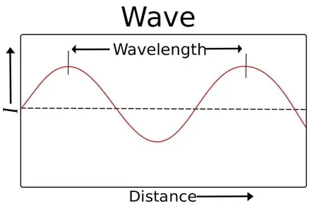
Although light exists as a particle, the way light travels is through an oscillating wave. The wavelength, as you would expect, is just the length. The length measures the distance between two crests in the wave. Visible light typically has a wavelength from about 400 to 700 nanometers.
What is Amplitude?
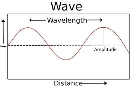
Amplitude is another measurement of the wave. Amplitude measures the height of the wave starting from the point of rest to the crest of the wave. What this measurement tells us practically, is the intensity of light or the brightness of light. So, a larger amplitude or a tall crest means a brighter or more intense light.
What is a Light Wave Phase?
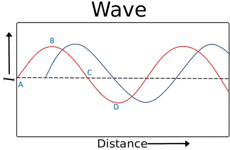
As we saw above, light travels in a wave and if you were to just look at the wave you would see it starts from the resting point or equilibrium point (dotted lines), goes up and reaches a crest, then a trough, then back to the equilibrium point.
This movement from A all the way to B is called a cycle. Within the cycle you could look at any given point and determine the phase at which the wave is in. These positions are measured in degrees kind of like a circle. For example from A to B would be 360 degrees.

If we look at the image below, we can see that point B is out of phase relative to point A 90 degrees and point A is out of phase relative to point C 180 degrees. Now imagine if we overlayered another distinct wave on top of this wave. You could look at the two waves and determine the degree to which the two waves were out of phase at any given point on those two waves. Keep this in mind for later.
Phase Contrast Components
There are two key components that enable us to manipulate the light in such a way to achieve the phase contrast result. There are some microscopes that are built specifically for phase contrast microscopy which will have these components already built in but if you have a standard brightfield microscope you can, with the right components, turn your microscope into a phase contrast microscope. This kit on Amazon that has everything you need to convert your microscope into a phase contrast microscope.
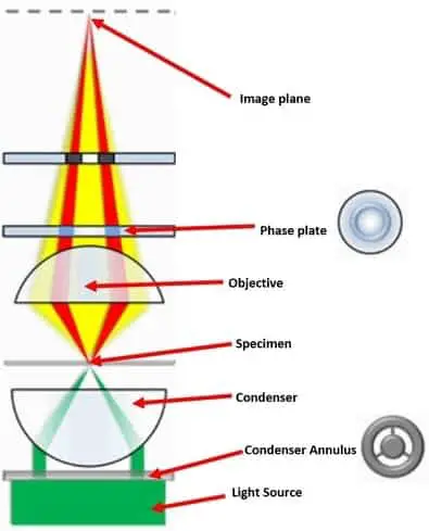
The first component is the phase contrast condenser with condenser annulus. This component is a specialized condenser meant specifically for phase contrast microscopy. The condenser contains a phase annulus which has flat black light absorbing plate with transparent annular rings.
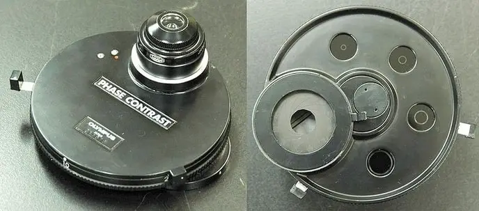
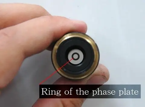
The next component is a phase contrast objective(s). These specialized objectives are built with a phase plate that works in conjunction with the condenser annulus to achieve the phase shift required for phase contrast microscopy. These types of objectives have a phase plate built into the rear of the objective. If you are looking for phase contrast objectives there are some on Amazon that are sometimes lower than the manufacturer website.
The phase plate can be either positive or negative, raised or lowered plates, to produce positive or negative phase contrast. Phase contrast objectives are sold as individual objectives but typically they are bought in a set containing multiple magnifications.
How Does Phase Contrast Work?
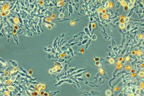
One of the most important parts of this process is the distinction between the diffracted and un-diffracted light that emerges from the specimen. Typically phase contrast is used on highly transparent specimens that do not absorb much light.
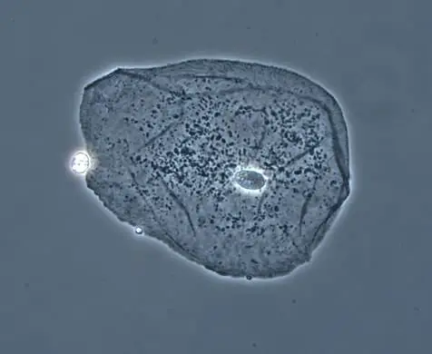
These types of specimens are called phase objects as opposed to amplitude objects that do absorb light. Some examples of these types of specimens are bacteria, tissue samples, cells, and other transparent microorganisms. Phase objects appear almost invisible under brightfield microscopy because the specimen diffracts light but does not absorb much light keeping the amplitude of light similar to the un-diffracted light.
The result is that the diffracted light is out of step by about ¼ of a wavelength with the un-diffracted light. Our eyes are not able to pick up details with this organization of light waves because of the lack of contrast. The object of the phase contrast technique is to slow the un-diffracted light by a ¼ of a wavelength making the waves further out of phase by a net difference of ½ a wavelength. With this ½ wavelength difference the human eye is able to see much more contrast on the image produced by the microscope.
The process starts with the phase contrast condenser where light is focused on the annulus which produces a hollow cone of light loosely speaking. Waves that pass through the annulus light the specimen with either diffracted or un-diffracted light. Diffracted light passing through the specimen is slowed down by structures in the specimen by about a ¼ wavelength.
Then the phase plate in the objective lens then slows down the un-diffracted waves. The resulting light that is focused on the image plane goes through either constructive or destructive interference which has the effect of increasing or decreasing the brightness of the specimen against the background light. The result is stark contrast, which enables the human eye to pick up on much more detail that originally could not be seen.
Advantages and Disadvantages of Phase Contrast
Advantages
- There are so many microorganisms that are transparent and thus would be a great candidate for using phase contrast.
- Phase contrast can give you hidden details and give you a sense of the density of the specimen which can be extremely helpful when looking at different cell organelles and cellular structures.
- Prior to phase contrast microscopy specimens would need to be stained or otherwise manipulated in order be seen under a standard brightfield microscope. This would have the obvious effect of either killing the specimen or contaminating it at the least. Phase contrast allows us to see freely moving, alive, and uncontaminated specimens with great detail.
Disadvantages
- Objectives in general are pretty pricy but specialized objectives like phase contrast objectives can quickly approach or exceed a $1,000 for a set of phase contrast objectives with a phase condenser depending on many factors and features. In some cases, for hobbyists this is prohibitively expensive unless you are really going to get some use and utility from it.
- A halo light ring is a natural result of using phase contrast, but most people consider this a distortionary effect that takes away from the image quality. You can see the halo light ring around the edges of the specimen and larger specimen features which typically will have a lighter shadow that resembles a glow similar to a halo. Some highly advanced phase contrast microscopes have corrected for this distortion using an external interferometric module.
History of Phase Contrast
Prior to the discovery of phase contrast, scientists had to resort to staining, using darkfield and oblique illumination techniques, and by defocusing the microscope. All of which had extreme downsides to visualizing highly transparent specimens.
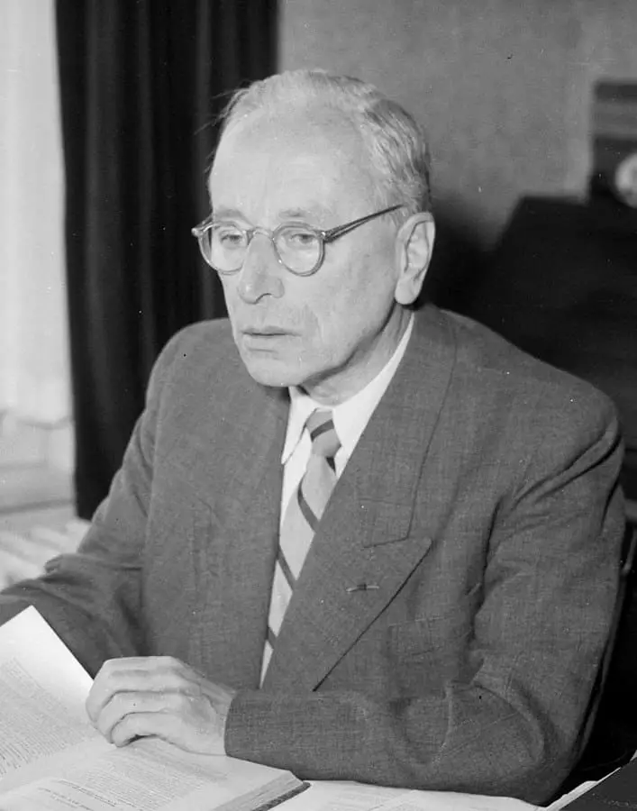
In 1934 Frits Zernike, a Dutch physicist, was studying spectral lines which eventually led him to discover specific behaviors of diffracted light with phase shifts. Zernkie applied what he discovered to microscopy leading to the invention of the phase contrast microscope during World War II. Zernkie eventually won a Nobel Prize for this amazing discovery and particularly the application of it to microscopy.
Since then phase contrast components have been refined and specialized with phase plates that have differing levels of light absorption to achieve the exact level of phase contrast required by the specimen and/or processes under observation. Today phase contrast objectives and condensers can be purchase at a relatively cheap price given the level of sophistication and what the components allow you to achieve on the microscope.
Takeaways
Phase contrast microscopy is an amazing example of using theoretical physics and applying it to solve real problems scientists were facing in the field of microbiology. This crucial technique has helped scientist expand their field of inquiry for many decades after it was invented. Phase contrast microscopy allows scientists to visualize specific organelles directly under a microscope without staining or otherwise contaminating the specimen.
Because of the hands off approach that phase contrast allows, scientists have been able to observe cellular functions and processes that many phase object microorganisms undergo that were previously very difficult or impossible to see. Phase contrast has also played an integral role in diagnosing tumors by visualizing, studying growth, dynamics, and behavior.
Phase contrast has been an amazing technique for science, and it blows my mind that we, as microscope hobbyists, can use the same techniques and see the same things that scientists see during intricate and complex experiments and observations.
References
- https://bio.libretexts.org/Bookshelves/Microbiology/Book%3A_Microbiology_(Boundless)/3%3A_Microscopy/3.3%3A_Other_Types_of_Microscopy/3.3B%3A_Phase-Contrast_Microscopy
- http://microscopy.berkeley.edu/Resources/instruction/phase_contrast.html
- https://www.nature.com/articles/srep44034
- https://www.researchgate.net/profile/Baoli_Yao/publication/51765403_Phase-shifting_Zernike_phase_contrast_microscopy_for_quantitative_phase_measurement/links/54c65e590cf2911c7a58bff2/Phase-shifting-Zernike-phase-contrast-microscopy-for-quantitative-phase-measurement.pdf
- http://www.qrg.northwestern.edu/projects/vss/docs/Communications/1-what-is-wavelength.html
- https://www.microscopyu.com/techniques/phase-contrast/introduction-to-phase-contrast-microscopy

![Duinen, […] van / Anefo](https://commons.wikimedia.org/wiki/File:Frits_Zernike_1953b.jpg)