

I was watching the magic school bus the other day with my daughter. It was the episode where the Ms. Frizzle and the class go into the human body. As a kid I loved the part where they are riding the bloodstream with the visible blood cells.
As an adult it gave me the idea to take a look at blood under my own microscope, after I cut my finger in the kitchen. It turns out that it is a very different experience when you are looking at your own blood cells and seeing them move and change shape right in front of your eyes.
In this article I’m going to go through the steps to setup this little experiment, and how you can do it yourself at home if you want to give it a try.
How to Get the Sample
In my case I already had a cut on my finger, but I would not suggest cutting yourself for this experiment. Instead I would use an ultra-fine pen needle that you can find at your local drug store for around $8 or $9. You can then safely and cleanly prick your finger to draw an extremely small amount of blood. You don’t need a lot for this lab don’t try to squeeze the wound or anything to get more blood out. A drop of blood is all you need.
Please be sure to read and follow the medical directions and best practices described on the box. Once you have mounted the slide, which we will go over in the next section, apply standard medical treatment to your wound site and ensure you have properly cleaned and bandaged the wound to prevent infection.
How to Mount the Slide
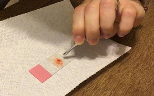
Once you’ve got a droplet of blood, we need to get it on a clean microscope slide. You can do this by landing the droplet of blood directly on the slide. When you place the blood droplet directly on the slide from your finger, make sure you do not touch your finger to the slide, only the blood. Otherwise you will get fingerprints on the slide which will reduce the quality of the image.
Next I would recommend adding a droplet of water on the blood sample so that you can see more of the blood cell movement. If you do not add water, the blood cells are usually quite close together and you will not get a very good image.
Now we need to cover the sample with a slide coverslip. To add the slide coverslip, you can use your fingers but if you have a set of tweezers handy, I would recommend using that to avoid fingerprints on the coverslip. Drop one side of the coverslip over the sample and then slowly lower the other side of the coverslip. If you drop the coverslip over the sample, you will most likely get air bubbles that will interfere with a clear image.
The next step is to lower the stage to the lowest level using the coarse focus adjustment knobs and place the slide over the condenser lens. Engage the stage clip and we are ready to go.
Best Way to Observe Blood Under the Microscope
To see the distinctive red blood cell disk shape, you need a little bit of contrast. I did this by lowering the condenser lens and closing the iris diaphragm to match the 0.65 numerical aperture of the 40X objective I was using. I did not use darkfield in my particular example but viewing red blood cells under a darkfield configuration is a very popular choice. Also using the phase contrast configuration will give you excellent contrast to see the disk shape of the red blood cell. The microscope I am using is this OMAX trinocular microscope. However I recently purchased this DSLR camera and hooked it up to my trinocular mound using this adapter and it takes much better pictures and video.
I was able to the see the disk shape under brightfield so I did not opt for these configurations but if you are having trouble seeing the disk shape you may want to try these options. I was not able to identify platelet cells, which are much smaller than red blood cells. I also was not able to identify white blood cells. This is either because I missed them or because they are less common in any given sample of blood.
Some Quick Information on Blood
Blood is a liquid made of mostly of plasma and cells but also contains protein which makes it thicker than water. Blood’s primary purpose is to circulate oxygen throughout the body, but it also helps to fight infections, clots wounds to stop bleeding, transports waste materials and chemicals the body’s filtration organs like the liver and kidney and helps keep the human body temperature constant.
Blood is composed mainly of the following materials:
- Red blood cells (erythrocyte)
- White blood cells (leukocytes)
- Platelets (thrombocytes)
- Plasma
A little over half of the composition of blood is plasma. Plasma is mostly water but there are other components like electrolytes, vitamins, glucose, and proteins that make up a smaller percentage of plasma.

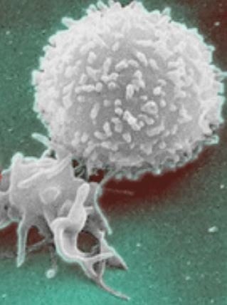
White blood cells are also present in blood and are a large part of your body’s overall immune response to things like harmful bacteria, and viruses. White bloods cells detect and actually eat or destroy harmful organisms with digestive enzymes.
The primary function of platelets is clotting when the body experiences a wound. The platelets pile on top of one another at the site of a wound and with the help of some red blood cells joining the pile they eventually clot the wound enabling the body to heal. During the healing process some of the platelets fall away and eventually when the wound is fully healed, the remainder of the platelets fall away from the site where the wound once was.

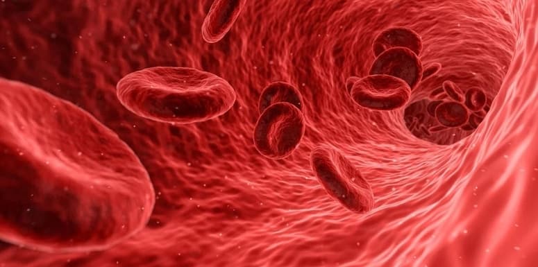
Red blood cells are responsible for transporting oxygen throughout the body. It also picks up carbon dioxide and transports it to the lungs for exhalation from the body. Red blood cells are the most common cells found in blood and is part of what gives blood the red color.
Anatomy of a Red Blood Cell
The only cell we’ve seen during inspection of the blood has been the red blood cell. This is primarily because it is the most common cell found in any given sample of blood. Let’s take a look at the anatomy of a cell and see what exactly makes up these little red blood cells.
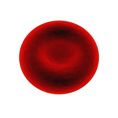
A red blood cell also called an erythrocyte, are disk shaped cells with a slightly concaved core. Similar to a donut except there is no hole. Red blood cells are about 6 micrometers in diameter and 2 micrometers thick making them one of the smallest cells in the entire body.
Red Blood cells are made primarily of hemoglobin molecules which binds to oxygen and is how red blood cells carry oxygen throughout the body. Red blood cells can maneuver and fit through some of the smallest veins and capillaries in the human body which we saw when we observed the red blood cells floating along with the water.
Although we call them cells they cannot replicate and do not have their own nucleus, or DNA. They also cannot express genes and synthesize proteins. Red blood cells only have a life span of about 4 months, so your bone marrow is constantly producing new red blood cells to replace the old ones.
Clean Up
After we’re done inspecting the blood under the microscope it is time to clean up.
- First, we need to take the blood sample slide off the microscope stage by lowering the stage, disengaging the stage clip(s) and placing it safely aside.
- Next we will lower the light intensity and turn off the illuminator.
- Unplug the microscope.
- Place the microscope cover over the microscope and put it back where it is stored.
- Next we need to clean the slide. The slide can be rinsed in a sink under cool water until the blood is removed from the slide.
- Next apply the cleaning solution to the slide and using lens paper wipe the slide until dry.
That’s it! Everything is clean and put away, ready for you next lab or experiment.
Takeaways
This was a pretty cool little lab. I’ve seen renditions, drawings, simulations, and video, of blood under a microscope but it really is a different experience looking at blood under a microscope when it is right in front of you and you can see the fluid dynamics and movements of the blood cells clearly.
It puts things in perspective when you realize that there were millions of red blood cells in that one drop of blood and our body contains around 1.5 gallons of blood!